File:Kollmann342.jpg: Difference between revisions
({{Kollmann1907}}) |
No edit summary |
||
| Line 1: | Line 1: | ||
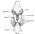
==Fig. 342. The anterior end of an embryo of Callorhynchus antarcticus (Shark)== | |||
viewed from the ventral side. (Norma frontal is). (According to Schauinsland.) | |||
The composition of the vertebrate head shape indicates a low-lying are identical in the main parts of the higher forms of development: the front part of the skull with frontal process, the nasal pits, the eye, through the pharyngeal membrane bay mouth closed, bounded on both sides by the mandibular arch (mandibular arch). Side come from the occipital the gill arch to unite in the middle. The second of which is commonly referred to as the hyoid arch. The gill filaments are dotted. The heart is in the range of the branchial arches as in the human embryo. | |||
{{GoogleTranslate}} | |||
{{Kollmann1907}} | {{Kollmann1907}} | ||
Fig. 342. Das vordere Körperende eines Embryo von Callorhynchus antarcticus (Haifisch) | |||
von der ventralen Seite betrachtet. (Norma frontalis.) | |||
(Nach Schauinsland.) | |||
Die Zusammensetzung des Wirbeltierkopfes einer tiefstehenden Form zeigt | |||
sich in den Hauptteilen identisch mit dem der höheren Entwicklungsformen: | |||
der Stirnteil des Schädels mit Stirnfortsatz, die Nasengruben, das Auge, die durch | |||
eine Rachenmembran geschlossene Mundbucht, zu beiden Seiten begrenzt durch | |||
die Unterkieferbogen (Arcus mandibularis). Seitlich kommen vom Hinterkopf | |||
die Kiemenbogen um sich in der Mitte zu vereinigen. Der zweite davon wird | |||
gewöhnlich als Hyoidbogen bezeichnet. Die Kiemenfäden sind punktiert. Das | |||
Herz liegt im Bereich der Kiemenbogen wie bei dem menschlichen Embryo. | |||
Revision as of 13:03, 17 October 2011
Fig. 342. The anterior end of an embryo of Callorhynchus antarcticus (Shark)
viewed from the ventral side. (Norma frontal is). (According to Schauinsland.)
The composition of the vertebrate head shape indicates a low-lying are identical in the main parts of the higher forms of development: the front part of the skull with frontal process, the nasal pits, the eye, through the pharyngeal membrane bay mouth closed, bounded on both sides by the mandibular arch (mandibular arch). Side come from the occipital the gill arch to unite in the middle. The second of which is commonly referred to as the hyoid arch. The gill filaments are dotted. The heart is in the range of the branchial arches as in the human embryo.
- This text is a Google translate computer generated translation and may contain many errors.
- This text is a Google translate computer generated translation and may contain many errors.
Images from - Atlas of the Development of Man (Volume 2)
(Handatlas der entwicklungsgeschichte des menschen)
- Kollmann Atlas 2: Gastrointestinal | Respiratory | Urogenital | Cardiovascular | Neural | Integumentary | Smell | Vision | Hearing | Kollmann Atlas 1 | Kollmann Atlas 2 | Julius Kollmann
- Links: Julius Kollman | Atlas Vol.1 | Atlas Vol.2 | Embryology History
| Historic Disclaimer - information about historic embryology pages |
|---|
| Pages where the terms "Historic" (textbooks, papers, people, recommendations) appear on this site, and sections within pages where this disclaimer appears, indicate that the content and scientific understanding are specific to the time of publication. This means that while some scientific descriptions are still accurate, the terminology and interpretation of the developmental mechanisms reflect the understanding at the time of original publication and those of the preceding periods, these terms, interpretations and recommendations may not reflect our current scientific understanding. (More? Embryology History | Historic Embryology Papers) |
Reference
Kollmann JKE. Atlas of the Development of Man (Handatlas der entwicklungsgeschichte des menschen). (1907) Vol.1 and Vol. 2. Jena, Gustav Fischer. (1898).
Cite this page: Hill, M.A. (2024, April 19) Embryology Kollmann342.jpg. Retrieved from https://embryology.med.unsw.edu.au/embryology/index.php/File:Kollmann342.jpg
- © Dr Mark Hill 2024, UNSW Embryology ISBN: 978 0 7334 2609 4 - UNSW CRICOS Provider Code No. 00098G
Fig. 342. Das vordere Körperende eines Embryo von Callorhynchus antarcticus (Haifisch)
von der ventralen Seite betrachtet. (Norma frontalis.)
(Nach Schauinsland.)
Die Zusammensetzung des Wirbeltierkopfes einer tiefstehenden Form zeigt
sich in den Hauptteilen identisch mit dem der höheren Entwicklungsformen:
der Stirnteil des Schädels mit Stirnfortsatz, die Nasengruben, das Auge, die durch
eine Rachenmembran geschlossene Mundbucht, zu beiden Seiten begrenzt durch
die Unterkieferbogen (Arcus mandibularis). Seitlich kommen vom Hinterkopf
die Kiemenbogen um sich in der Mitte zu vereinigen. Der zweite davon wird
gewöhnlich als Hyoidbogen bezeichnet. Die Kiemenfäden sind punktiert. Das
Herz liegt im Bereich der Kiemenbogen wie bei dem menschlichen Embryo.
File history
Click on a date/time to view the file as it appeared at that time.
| Date/Time | Thumbnail | Dimensions | User | Comment | |
|---|---|---|---|---|---|
| current | 12:54, 16 October 2011 |  | 728 × 699 (59 KB) | S8600021 (talk | contribs) | {{Kollmann1907}} |
You cannot overwrite this file.
File usage
The following 2 pages use this file:
