File:Kollmann061.jpg

Original file (1,000 × 512 pixels, file size: 78 KB, MIME type: image/jpeg)
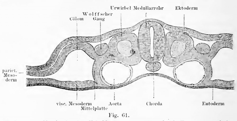
Fig. 61. Chicken embryo 4 somites from the beginning of third day
cross-section. Magnified 170 times
(Fig. 61), which passes into the ventral walls of the trunk. The other sheet follows the endoderm, mesoderm, and is called thevisceral layer 2). Ans It will make the muscular wall and serosa of the intestine, as well as the mesenteries. The column itself expands and fills Bichmitllrlymphe. It is called primitive body cavity or coelom: the first stage of the laterPleuroperitonealhöhle which intestinal tube and its appendages throughout thevertebrate kingdom absorbs. The coelom does not extend into the root zone (Fig.Gl), and not in the front section of the Parietalzone which involved a look at theformation of the head. The splitting of the mesoderm has recently been seen in a human embryo, its mass Fruchthof 1.54 mm in length: its age was about 10 days.The column starts at the peri-phery of the fruit farm and is just going into thesolid-parietal
- This text is a Google translate computer generated translation and may contain many errors.
Images from - Atlas of the Development of Man (Volume 1)
(Handatlas der entwicklungsgeschichte des menschen)
- Kollmann Atlas 1: Predevelopment | Ontogeny | Fetal membranes | Body shape | Systems and organs | Kollmann Atlas 1 | Kollmann Atlas 2 | Julius Kollmann
- Links: Julius Kollman | Atlas Vol.1 | Atlas Vol.2 | Embryology History
| Historic Disclaimer - information about historic embryology pages |
|---|
| Pages where the terms "Historic" (textbooks, papers, people, recommendations) appear on this site, and sections within pages where this disclaimer appears, indicate that the content and scientific understanding are specific to the time of publication. This means that while some scientific descriptions are still accurate, the terminology and interpretation of the developmental mechanisms reflect the understanding at the time of original publication and those of the preceding periods, these terms, interpretations and recommendations may not reflect our current scientific understanding. (More? Embryology History | Historic Embryology Papers) |
Fig. 61. Embryo vom Vogel mit vier Keimblättern vom Anfang des 3. Tages, Querschnitt. 170 mal vergr. (Fig. 61), das in die ventralen Wandungen des Rumpfes übergeht. Das andere Blatt folgt dem Entoderm und heisst viscerales Blatt des Mesoderm 2 ). Ans ihm geht die muskulöse Wand und die Serosa des Darmrohres hervor, ebenso die Mesenterien. Die Spalte selbst erweitert sich und füllt BichmitUrlymphe. Sie heisst primitive Leibeshöhle oder Cölom: die erste Stufe der späteren Pleuroperitonealhöhle, welche Darm- rohr und seine Adnexa durch das ganze Wirbeltierreich aufnimmt. Das Cölom erstreckt sich nicht in die Stammzone hinein (Fig. Gl), und nicht in den vorderen Abschnitt der Parietalzone, die sieh an der Bildung des Kopfes beteiligt. Die Spaltung des Mesoderm ist jüngst bei einem menschlichen Embryo gesehen worden; sein Fruchthof mass in der Länge 1,54 mm: sein Alter war ca. 10 Tage. Die Spalte beginnt an der Peri- pherie des Fruchthofes und ist eben im Begriff, in die solide Parietal-
File history
Click on a date/time to view the file as it appeared at that time.
| Date/Time | Thumbnail | Dimensions | User | Comment | |
|---|---|---|---|---|---|
| current | 15:53, 30 October 2011 |  | 1,000 × 512 (78 KB) | S8600021 (talk | contribs) | ==Fig. 61. == {{Kollmann1906}} Category:Mesoderm |
You cannot overwrite this file.
File usage
The following page uses this file:
