File:Kingsbury1932 fig1.jpg: Difference between revisions

Original file (1,000 × 1,047 pixels, file size: 113 KB, MIME type: image/jpeg)
mNo edit summary |
mNo edit summary |
||
| Line 1: | Line 1: | ||
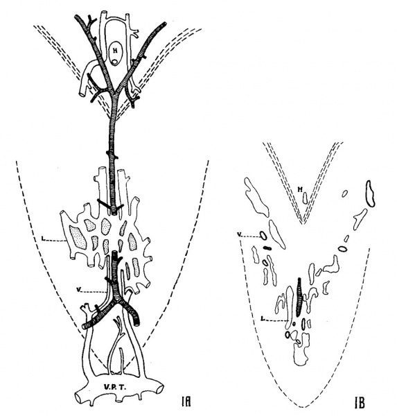
Fig. 1 A. Flat reconstruction (semidiagrammatic) made from a. series through the pharyngeal tonsil of a cat fetus, 100-mm. length, cut in a plane parallel to the surface. Arteries cross-marked; veins (1)) with heavy outline; basal lymphatic channels (L) shown by light line (diagrammatic). V.P.1'., transverse pharyngeal vein; H, hypophyseal stalk. Caudal end is down. Boundary of the pharyngeal region is indicated by broken line. X 20. B. Reproduction of a single section from the series (100-mm. fetu), correctly plotting vascular channels. Glands and developing mesenchymal tissue not indicated. X 20. | ==Fig. 1 == | ||
A. Flat reconstruction (semidiagrammatic) made from a. series through the pharyngeal tonsil of a cat fetus, 100-mm. length, cut in a plane parallel to the surface. Arteries cross-marked; veins (1)) with heavy outline; basal lymphatic channels (L) shown by light line (diagrammatic). V.P.1'., transverse pharyngeal vein; H, hypophyseal stalk. Caudal end is down. Boundary of the pharyngeal region is indicated by broken line. X 20. | |||
B. Reproduction of a single section from the series (100-mm. fetu), correctly plotting vascular channels. Glands and developing mesenchymal tissue not indicated. X 20. | |||
===Reference=== | ===Reference=== | ||
Latest revision as of 22:05, 28 March 2017
Fig. 1
A. Flat reconstruction (semidiagrammatic) made from a. series through the pharyngeal tonsil of a cat fetus, 100-mm. length, cut in a plane parallel to the surface. Arteries cross-marked; veins (1)) with heavy outline; basal lymphatic channels (L) shown by light line (diagrammatic). V.P.1'., transverse pharyngeal vein; H, hypophyseal stalk. Caudal end is down. Boundary of the pharyngeal region is indicated by broken line. X 20.
B. Reproduction of a single section from the series (100-mm. fetu), correctly plotting vascular channels. Glands and developing mesenchymal tissue not indicated. X 20.
Reference
Kingsbury BF. The developmental significance of the mammalian pharyngeal tonsil - Cat. (1932) Amer. J Anat. 50(2): 201-231.
Cite this page: Hill, M.A. (2024, April 19) Embryology Kingsbury1932 fig1.jpg. Retrieved from https://embryology.med.unsw.edu.au/embryology/index.php/File:Kingsbury1932_fig1.jpg
- © Dr Mark Hill 2024, UNSW Embryology ISBN: 978 0 7334 2609 4 - UNSW CRICOS Provider Code No. 00098G
File history
Click on a date/time to view the file as it appeared at that time.
| Date/Time | Thumbnail | Dimensions | User | Comment | |
|---|---|---|---|---|---|
| current | 22:04, 28 March 2017 |  | 1,000 × 1,047 (113 KB) | Z8600021 (talk | contribs) | |
| 22:04, 28 March 2017 |  | 1,351 × 1,676 (324 KB) | Z8600021 (talk | contribs) |
You cannot overwrite this file.
File usage
The following page uses this file: