File:Kingsbury1922 plate01.jpg
Original file (1,876 × 2,227 pixels, file size: 579 KB, MIME type: image/jpeg)
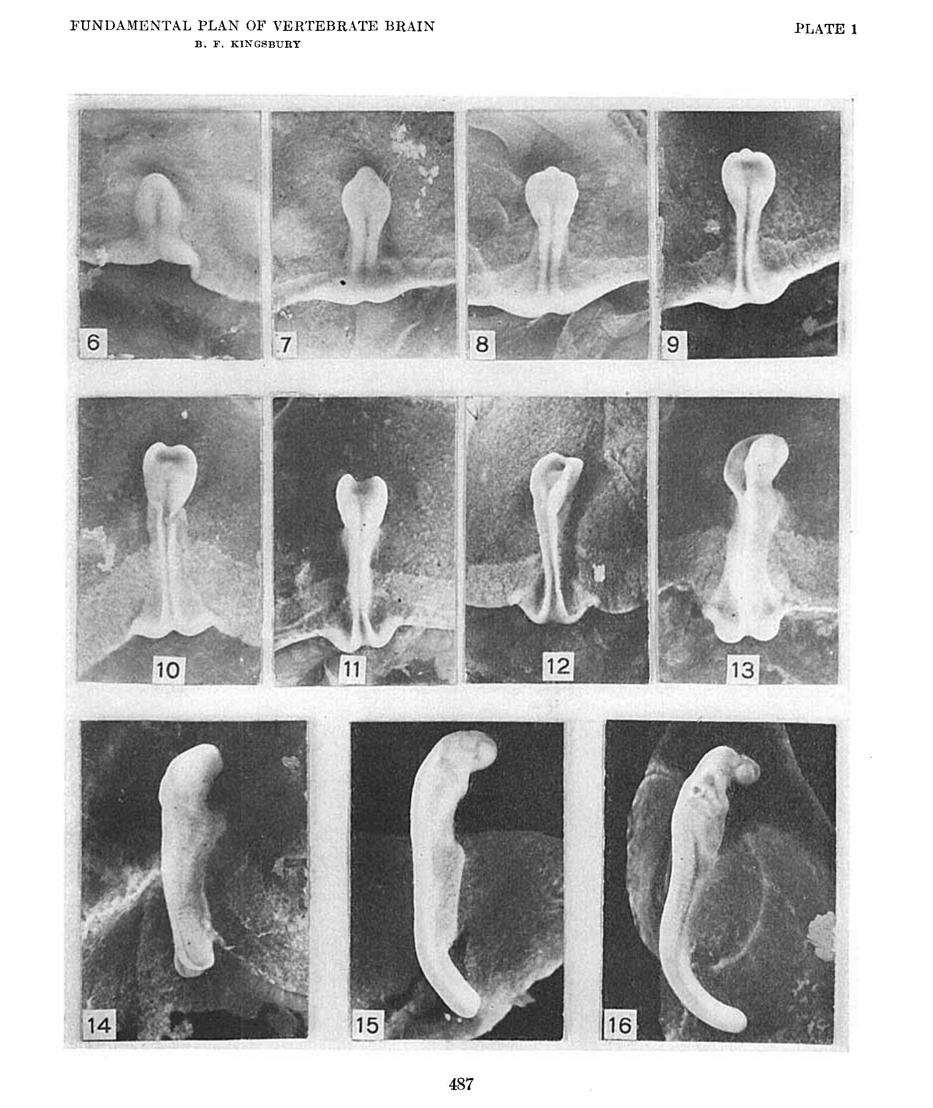
Plate 1
Photographs, all at a magnification of ten diameters, except figure 16 which is X 8. Embryos of Squalus acanthias dorsal aspect.
Online Editor - Squalus acanthias = Spiny dogfish
6. Series 42, no somites. length 1.5 mm.
7. Series 45, 6 to 7 somites, length 2.2 mm.
8. Series not sectioned, length 2.4 mm.
9. Series 48, ca. 8 somites, length 2.16 mm.
10. Series not sectioned, length 2.18 mm.
11. Series 52, 11 to 12 somites, length 2.9 mm.
12. Series 54, 13 to 14 somites, length 3.5 mm.
13. Series 55, 15 somites length, 4.0 mm.
14. Series 59, ca. 17 somites, length 4.8 mm.
15. Series 69, 29 somites, length 5.0+ mm.
16. Series 75, 41 somites, length ca. 7.0 mm.
Reference
Kingsbury BF. The fundamental plan of the vertebrate brain. (1922) J. Comp. Neural. 461-490.
Cite this page: Hill, M.A. (2024, April 23) Embryology Kingsbury1922 plate01.jpg. Retrieved from https://embryology.med.unsw.edu.au/embryology/index.php/File:Kingsbury1922_plate01.jpg
- © Dr Mark Hill 2024, UNSW Embryology ISBN: 978 0 7334 2609 4 - UNSW CRICOS Provider Code No. 00098G
File history
Click on a date/time to view the file as it appeared at that time.
| Date/Time | Thumbnail | Dimensions | User | Comment | |
|---|---|---|---|---|---|
| current | 13:51, 22 November 2019 | | 1,876 × 2,227 (579 KB) | Z8600021 (talk | contribs) | {{Ref-Kingsbury1922}} |
You cannot overwrite this file.
File usage
The following page uses this file: