File:Kingsbury1922 fig05.jpg

Original file (1,353 × 1,412 pixels, file size: 302 KB, MIME type: image/jpeg)
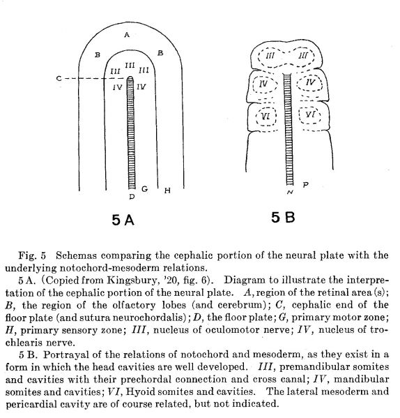
Fig. 5
Schemas comparing the cephalic portion of the neural plate with the underlying notochord—mesoderm relations.
5 A. (Copied from Kingsbury, ’20, fig. 6). Diagram to illustrate the interpretation of the cephalic portion of the neural plate. A, region of the retinal area (s); B, the region of the olfactory lobes (and cerebrum); C, cephalic end of the floor plate (and sutura neurochordalis) ,' D, the floor plate; G, primary motor zone; H, primary sensory zone; III, nucleus of oculomotor nerve; IV, nucleus of trochlearis nerve. '
5 B. Portrayal of the relations of notochord and mesoderm, as they exist in a form in which the head cavities are well developed. III, premandibular somites and cavities with their prechordal connection and cross canal; IV, mandibular somites and cavities; VI, Hyoid somites and cavities. The lateral mesoderm and pericardial cavity are of course related, but not indicated.
Reference
Kingsbury BF. The fundamental plan of the vertebrate brain. (1922) J. Comp. Neural. 461-490.
Cite this page: Hill, M.A. (2024, April 19) Embryology Kingsbury1922 fig05.jpg. Retrieved from https://embryology.med.unsw.edu.au/embryology/index.php/File:Kingsbury1922_fig05.jpg
- © Dr Mark Hill 2024, UNSW Embryology ISBN: 978 0 7334 2609 4 - UNSW CRICOS Provider Code No. 00098G
File history
Click on a date/time to view the file as it appeared at that time.
| Date/Time | Thumbnail | Dimensions | User | Comment | |
|---|---|---|---|---|---|
| current | 10:09, 23 November 2019 | | 1,353 × 1,412 (302 KB) | Z8600021 (talk | contribs) |
You cannot overwrite this file.
File usage
The following page uses this file: