File:Keith1921 fig021.jpg
Original file (1,179 × 625 pixels, file size: 160 KB, MIME type: image/jpeg)
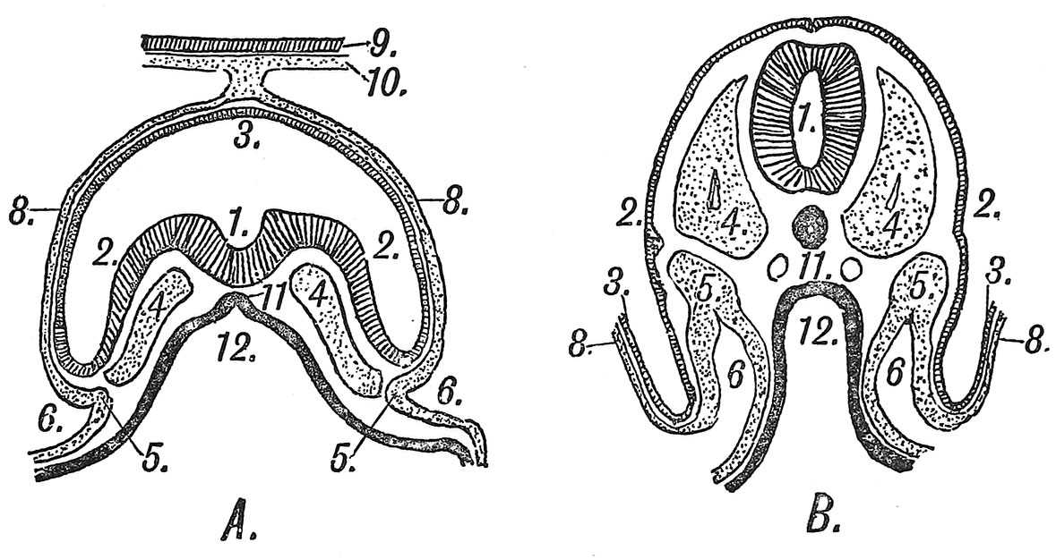
Fig. 21. Schematic Transverse Sections of two Human Embryos
| A In the 3rd. week of development. | B In the 4th week of development. |
The numbers are placed on corresponding points : Epiblast, shaded ; hypoblast, black ; mesoblast, stippled.
| 1. Neural groove and canal.
2. Epiblast of embryo. 3. Epiblast lining amnion. Only the attachment of the amnion is represented in B. 4. Paraxial mesoblast. 5. Intermediate cell mass. |
6. Coelom, bounded by the somatopleure externally and splanchnopleure internally.
8. Mesoblast on amnion. 9, 10. Chorion. 11. Notochord. 12. Archenteron. |
- Chapter 1 Figures: 1 | 2 | 3 | 4 | 5 | 6 | 7 | 8 | 9 | 10 | 11 | 12 | 13 | 14 | 15 | 16 | 17 | 18 | 19 | 20 | 21 | 22 | 23 | All Figures
| Historic Disclaimer - information about historic embryology pages |
|---|
 |
Human Embryology and Morphology: 1 Early Ovum and Embryo | 2 Connection between Foetus and Uterus | 3 Primitive Streak Notochord and Somites | 4 Age Changes | 5 Spinal Column and Back | 6 Body Segmentation | 7 Spinal Cord | 8 Mid- and Hind-Brains | 9 Fore-Brain | 10 Fore-Brain Cerebral Vesicles | 11 Cranium | 12 Face | 13 Teeth and Mastication | 14 Nasal and Olfactory | 15 Sense OF Sight | 16 Hearing | 17 Pharynx and Neck | 18 Tongue, Thyroid and Pharynx | 19 Organs of Digestion | 20 Circulatory System | 21 Circulatory System (continued) | 22 Respiratory System | 23 Urogenital System | 24 Urogenital System (Continued) | 25 Body Wall and Pelvic Floor | 26 Limb Buds | 27 Limbs | 28 Skin and Appendages | Figures
Reference
Keith A. Human Embryology and Morphology. (1921) New York, Longmans, Green & Co. London: Edward Arnold.
Cite this page: Hill, M.A. (2024, April 19) Embryology Keith1921 fig021.jpg. Retrieved from https://embryology.med.unsw.edu.au/embryology/index.php/File:Keith1921_fig021.jpg
- © Dr Mark Hill 2024, UNSW Embryology ISBN: 978 0 7334 2609 4 - UNSW CRICOS Provider Code No. 00098G
File history
Click on a date/time to view the file as it appeared at that time.
| Date/Time | Thumbnail | Dimensions | User | Comment | |
|---|---|---|---|---|---|
| current | 10:08, 22 December 2014 | | 1,179 × 625 (160 KB) | Z8600021 (talk | contribs) | |
| 09:48, 22 December 2014 | 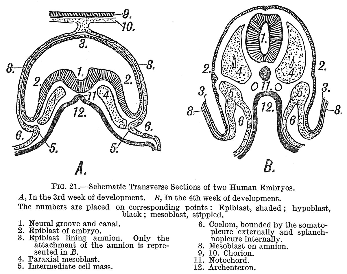 | 1,200 × 947 (254 KB) | Z8600021 (talk | contribs) |
You cannot overwrite this file.
File usage
The following 2 pages use this file: