File:Keith1902 fig213a.jpg

Original file (854 × 800 pixels, file size: 166 KB, MIME type: image/jpeg)
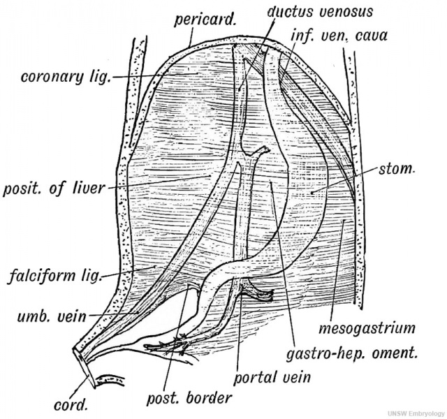
Fig. 213. The origin of the Peritoneal Ligaments connected with the Liver
When the liver grows out from the septum transversum it withdraws within the ventral mesentery. Out of the ventral mesentery are formed all the ligaments of the liver (Fig. 213 A). These are the following:
1. The Gastro-hepatic Omentum is that part of the ventral mesentery which passes from (1) the oesophagus, (2) lesser curvature or ventral border of stomach, and (3) first stage of duodenum to (1) the diaphragm, (2) the posterior part of the longitudinal fissure of the liver, the ductus venosus lying within its hepatic attachment, and (3) the transverse fissure of the liver (Fig. 213 B). The portal and umbilical veins lie in the ventral mesentery (Fig. 213^4); the hepatic artery passes by it to the liver. The right or free border of the gastro-hepatic omentum, with the falciform ligament containing the remnant of the umbilical vein, represents the posterior border of the primitive ventral mesentery (Fig. 213 A).
2. The Falciform Ligament, containing the umbilical vein, also represents part of the ventral mesentery (Fig. 213 A). At an early stage the umbilical veins reached the sinus venosus by passing through the septum transversum. The development of the liver led to its being thrust out within the falciform ligament.
3. The coronary, the right and left lateral ligaments, and the attachments to the vena cava and diaphragm (Fig. 213 5) are developed as the liver emerges from the septum transversum, and separates itself from the diaphragm.
- Organs of Digestion: Fig. 212. Mesentery of the Fore-gut | Fig. 213 A. Liver Peritoneal Ligaments | Fig. 213 B. Liver Peritoneal Ligaments | Fig. 214. Mammalian Liver | Fig. 215. Liver of a human foetus 3rd month | Fig. 216. Embryo Spleen, Pancreas, and Liver to the Mesogastrium | Fig. 217. Mesogastrium | Fig. 218. Pancreatic and Hepatic Processes human embryo 4th week | Fig. 219. Arrangement of Vessels in the Dorsal week | Fig. 220. Formation of the Lesser Sac of the Peritoneum from the Dorsal Mesogastrium | Fig. 221. Alimentary Canal human embryo 3rd week | Fig. 222. Alimentary Canal 5th week | Fig. 223 A. The mesentery of the hind-gut | Fig. 223 B. descending Meso-colon becomes applied to the parietal Peritoneum | Fig. 224. Apex of the Caecum at birth | Fig. 225 A. Appendix and Peritoneal Folds 2nd month | Fig. 225 B. Peritoneal Fossae in the lleo-caecal Region | Fig. 226. Rotation of the Intestinal Loop
| Historic Disclaimer - information about historic embryology pages |
|---|
 |
Human Embryology and Morphology (1902): Development or the Face | The Nasal Cavities and Olfactory Structures | Development of the Pharynx and Neck | Development of the Organ of Hearing | Development and Morphology of the Teeth | The Skin and its Appendages | The Development of the Ovum of the Foetus from the Ovum of the Mother | The Manner in which a Connection is Established between the Foetus and Uterus | The Uro-genital System | Formation of the Pubo-femoral Region, Pelvic Floor and Fascia | The Spinal Column and Back | The Segmentation of the Body | The Cranium | Development of the Structures concerned in the Sense of Sight | The Brain and Spinal Cord | Development of the Circulatory System | The Respiratory System | The Organs of Digestion | The Body Wall, Ribs, and Sternum | The Limbs | Figures | Embryology History
Reference
Keith A. Human Embryology and Morphology. (1902) London: Edward Arnold.
Cite this page: Hill, M.A. (2024, April 18) Embryology Keith1902 fig213a.jpg. Retrieved from https://embryology.med.unsw.edu.au/embryology/index.php/File:Keith1902_fig213a.jpg
- © Dr Mark Hill 2024, UNSW Embryology ISBN: 978 0 7334 2609 4 - UNSW CRICOS Provider Code No. 00098G
File history
Click on a date/time to view the file as it appeared at that time.
| Date/Time | Thumbnail | Dimensions | User | Comment | |
|---|---|---|---|---|---|
| current | 19:09, 20 January 2014 | | 854 × 800 (166 KB) | Z8600021 (talk | contribs) |
You cannot overwrite this file.
File usage
The following 4 pages use this file: