File:Keith1902 fig173.jpg

Original file (800 × 608 pixels, file size: 96 KB, MIME type: image/jpeg)
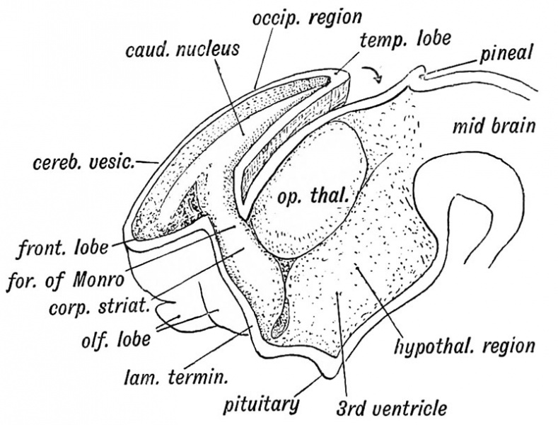
Fig. 173. Showing the Development of the Corpus Striatum in the floor and outer wall of the Cerebral Vesicle
The Corpus Striatum. — As soon as the cerebral vesicle grows it, the corpus striatum appears as a thickening in its wall at e outer side of the foramen of Monro (Fig. 173). With the isterior development of the vesicle it comes to lie in the venieular floor, from the foramen of Monro to its posterior tremity.
The anterior extremity of the corpus striatum is continuous ith the olfactory lobe (Fig. 173). Hence in the adult brain e anterior end of the corpus striatum appears at the base of the ain in the anterior perforated space, a lamina of grey matter rived from the olfactory lobe (Fig. 18, p. 22). As the cerebral side grows over the optic thalamus (Fig. 173) the corpus striatum comes to be applied to that body. The posterior extremity of the cerebral vesicle, in which the tail of the caudate nucleus is situated (Fig. 173), develops downwards, behind and below the optic thalamus, forming the descending horn of the lateral ventricle. Hence the occurrence of the tail of the caudate nucleus and the amygdaloid nucleus on the roof of the descending horn, both being intrinsic parts of the corpus striatum. The corpus striatum is imperfectly separated, during the 4th and 5th months of foetal life, into two parts, the caudate and lenticular nuclei, by the downgrowth, from the cerebral cortex of the fibres which form the internal capsule, crusta, and pyramidal tracts.
- Brain and Spinal Cord: Fig. 158. Human Neural Tube about 14 days | Fig. 159. Neural Tube Four Primary Divisions | Fig. 160. Zones of the Spinal Neural Tube Gth week | Fig. 161. Zones of the Embryonic Spinal Cord. | Fig. 162. Hind Brain 5th week | Fig. 163. Lateral Neural Tube 5th week | Fig. 164. Inferior Medullary Velum 5th month | Fig. 165. Frog Cerebellum and 4th Ventricle | Fig. 166. Human Cerebellum 3rd month | Fig. 167. walls of the fore-brain | Fig. 168. Human Cerebral Vesicles and Thalamencephalon 3rd month | Fig. 169. Adult 3rd Ventricle | Fig. 170. Fore and Mid-brain 5th week | Fig. 171. Fetal Brain 4th month | Fig. 172. Lamina Terminalis and Primitive Callosal Gyrus | Fig. 173. Corpus Striatum in Cerebral Vesicle | Fig. 174. Cerebral Hemisphere 2nd month | Fig. 175. Cerebral Hemisphere 5th month | Cerebral Hemisphere 7th month | Fiq. 177. Opercula and Fissure of Sylvius | Fig. 178. Ape Island of Reil and Fissures | Fig. 179A. Anthropoid Island of Reil | Fig. 180. Cranial Nerves and Primitive Segments | Figures
| Historic Disclaimer - information about historic embryology pages |
|---|
 |
Human Embryology and Morphology (1902): Development or the Face | The Nasal Cavities and Olfactory Structures | Development of the Pharynx and Neck | Development of the Organ of Hearing | Development and Morphology of the Teeth | The Skin and its Appendages | The Development of the Ovum of the Foetus from the Ovum of the Mother | The Manner in which a Connection is Established between the Foetus and Uterus | The Uro-genital System | Formation of the Pubo-femoral Region, Pelvic Floor and Fascia | The Spinal Column and Back | The Segmentation of the Body | The Cranium | Development of the Structures concerned in the Sense of Sight | The Brain and Spinal Cord | Development of the Circulatory System | The Respiratory System | The Organs of Digestion | The Body Wall, Ribs, and Sternum | The Limbs | Figures | Embryology History
Reference
Keith A. Human Embryology and Morphology. (1902) London: Edward Arnold.
Cite this page: Hill, M.A. (2024, April 18) Embryology Keith1902 fig173.jpg. Retrieved from https://embryology.med.unsw.edu.au/embryology/index.php/File:Keith1902_fig173.jpg
- © Dr Mark Hill 2024, UNSW Embryology ISBN: 978 0 7334 2609 4 - UNSW CRICOS Provider Code No. 00098G
File history
Click on a date/time to view the file as it appeared at that time.
| Date/Time | Thumbnail | Dimensions | User | Comment | |
|---|---|---|---|---|---|
| current | 11:31, 18 January 2014 | | 800 × 608 (96 KB) | Z8600021 (talk | contribs) |
You cannot overwrite this file.
File usage
The following 3 pages use this file: