File:Keith1902 fig080.jpg: Difference between revisions
mNo edit summary |
mNo edit summary |
||
| (2 intermediate revisions by the same user not shown) | |||
| Line 1: | Line 1: | ||
==Fig. 80. Diagrammatic section to show the position of the Wolffian and Genital Ridges on the dorsal wall of the abdomen== | ==Fig. 80. Diagrammatic section to show the position of the Wolffian and Genital Ridges on the dorsal wall of the abdomen== | ||
At the beginning of the second month of foetal life, the Wolffian body is well developed : by the end of that month it has become vestigial, the only parts remaining being those connected with the genital organs. It projects as a ridge from the lumbar and dorsal regions on each side of the mesentery, extending, on each side of the spine, from the posterior cervical region, where the diaphragm is developed, to the pelvis behind, where the ridges become approximated (Fig. 80). To its inner side, in the lower dorsal region, lies the genital ridge. The genital and the Wolffian bodies have each its own mesentery but these two mesenteries have a common attachment — the common uro-genital mesentery (Fig. 80). On section the Wolffian ridge is seen to be made up of convoluted tubules terminating at their blind extremities in glomeruli. The tubules open into the Wolffian duct just as in the frog; the duct is situated in the basal or attached part of the ridge. It runs backwards in this ridge and turns into the pelvis to end with the Müllerian duct (also situated in the Wolffian ridge) in the cloaca of the hind gut. | |||
{{Keith1902_9_figures}} | {{Keith1902_9_figures}} | ||
Latest revision as of 18:25, 22 January 2014
Fig. 80. Diagrammatic section to show the position of the Wolffian and Genital Ridges on the dorsal wall of the abdomen
At the beginning of the second month of foetal life, the Wolffian body is well developed : by the end of that month it has become vestigial, the only parts remaining being those connected with the genital organs. It projects as a ridge from the lumbar and dorsal regions on each side of the mesentery, extending, on each side of the spine, from the posterior cervical region, where the diaphragm is developed, to the pelvis behind, where the ridges become approximated (Fig. 80). To its inner side, in the lower dorsal region, lies the genital ridge. The genital and the Wolffian bodies have each its own mesentery but these two mesenteries have a common attachment — the common uro-genital mesentery (Fig. 80). On section the Wolffian ridge is seen to be made up of convoluted tubules terminating at their blind extremities in glomeruli. The tubules open into the Wolffian duct just as in the frog; the duct is situated in the basal or attached part of the ridge. It runs backwards in this ridge and turns into the pelvis to end with the Müllerian duct (also situated in the Wolffian ridge) in the cloaca of the hind gut.
| Historic Disclaimer - information about historic embryology pages |
|---|
 |
- The Uro-genital System: Fig. 79. Wolffian Body | Fig. 80. Wolffian and Genital Ridges | Fig. 81. Female Wolffian Body Remnants | Fig. 82. Male Wolffian Body Remnants |Fig. 83. Renal Bud | Fig. 84. Ureter in the Bladder | Fig. 85. Wolffian and Müllerian Ducts | Fig. 86. Genital Ducts 3rd month | Fig. 87. Müllerian Ducts 3rd month | Fig. 88. Uterus | Fig. 89. Uterus and Vagina | Fig. 90. Prostate remnants of Müllerian Ducts | Fig. 91. Prostate showing an unusual Uterus Masculinus | Fig. 92. Female Uro-genital Sinus | Fig. 93. Male Uro-genital Sinus | Fig. 94. Vagina and Uterus at 7th month | Fig. 95. Division of the Cloaca | Fig. 96. Imperforate Anus | Fig. 97. Cloacal Septum has failed to fuse with Perineal Septum | Fig. 98. The Uro-genital Cleft 2nd month | Fig. 99. Male bladder and urethra at birth | Fig. 100. Ectopia Vesicae | Fig. 101. Prostatic Tubules | Fig. 102. Testis in a foetus of 2.5 months | Fig. 103. Testis at the 6th month | Fig. 104. Inguinal Canal and Coverings of the Testis | Fig. 105. Processus Vaginalis | Figures
| Historic Disclaimer - information about historic embryology pages |
|---|
|
Human Embryology and Morphology (1902): Development or the Face | The Nasal Cavities and Olfactory Structures | Development of the Pharynx and Neck | Development of the Organ of Hearing | Development and Morphology of the Teeth | The Skin and its Appendages | The Development of the Ovum of the Foetus from the Ovum of the Mother | The Manner in which a Connection is Established between the Foetus and Uterus | The Uro-genital System | Formation of the Pubo-femoral Region, Pelvic Floor and Fascia | The Spinal Column and Back | The Segmentation of the Body | The Cranium | Development of the Structures concerned in the Sense of Sight | The Brain and Spinal Cord | Development of the Circulatory System | The Respiratory System | The Organs of Digestion | The Body Wall, Ribs, and Sternum | The Limbs | Figures | Embryology History
Reference
Keith A. Human Embryology and Morphology. (1902) London: Edward Arnold.
Cite this page: Hill, M.A. (2024, April 18) Embryology Keith1902 fig080.jpg. Retrieved from https://embryology.med.unsw.edu.au/embryology/index.php/File:Keith1902_fig080.jpg
- © Dr Mark Hill 2024, UNSW Embryology ISBN: 978 0 7334 2609 4 - UNSW CRICOS Provider Code No. 00098G
File history
Click on a date/time to view the file as it appeared at that time.
| Date/Time | Thumbnail | Dimensions | User | Comment | |
|---|---|---|---|---|---|
| current | 10:26, 7 January 2014 | 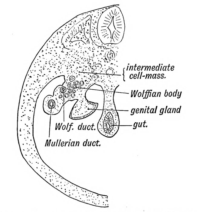 | 651 × 700 (79 KB) | Z8600021 (talk | contribs) |
You cannot overwrite this file.
File usage
The following 5 pages use this file: