File:Keith1902 fig079.jpg: Difference between revisions
mNo edit summary |
|||
| (One intermediate revision by the same user not shown) | |||
| Line 1: | Line 1: | ||
==Fig. 79. Scheme of the Wolffian Body of the right side== | |||
In Fig. 79 the Wolffian body, such as occurs in the frog, is represented diagrammatically and it corresponds in structure to the Wolffian body which appears in the human embryo. Each body is made up of a main duct and a series of tubules. In the frog, as in the human embryo, the hind gut ends in a dilatation, the cloaca. In the cloaca open the rectum, allantois, and the two Wolffian ducts — right and left. In the frog, the Wolffian bodies lie on each side of the spine, their anterior ends reaching forwards to the region of the heart. Each duct is joined by numerous convoluted tubules — the Wolffian tubules. Each tubule is furnished with a glomerulus at its blind extremity and in most features agrees with a secretory tubule — such as are seen in the permanent kidney. These tubules secrete the urine ; the Wolffian duct conveys the urine from the tubules to the cloaca. The anterior tubules, however, loose their secretory function and become associated with the genital gland. In the male frog they convey the spermatozoa to the Wolffian duct, which thus carries both urine, and spermatozoa. In the female, the genital Wolffian tubules are connected with the ovary but are quite functionless (Fig. 79). | |||
{{Historic Disclaimer}} | |||
{{Keith1902_9_figures}} | {{Keith1902_9_figures}} | ||
{{Human embryology morphology 1902 footer}} | {{Human embryology morphology 1902 footer}} | ||
Latest revision as of 18:22, 22 January 2014
Fig. 79. Scheme of the Wolffian Body of the right side
In Fig. 79 the Wolffian body, such as occurs in the frog, is represented diagrammatically and it corresponds in structure to the Wolffian body which appears in the human embryo. Each body is made up of a main duct and a series of tubules. In the frog, as in the human embryo, the hind gut ends in a dilatation, the cloaca. In the cloaca open the rectum, allantois, and the two Wolffian ducts — right and left. In the frog, the Wolffian bodies lie on each side of the spine, their anterior ends reaching forwards to the region of the heart. Each duct is joined by numerous convoluted tubules — the Wolffian tubules. Each tubule is furnished with a glomerulus at its blind extremity and in most features agrees with a secretory tubule — such as are seen in the permanent kidney. These tubules secrete the urine ; the Wolffian duct conveys the urine from the tubules to the cloaca. The anterior tubules, however, loose their secretory function and become associated with the genital gland. In the male frog they convey the spermatozoa to the Wolffian duct, which thus carries both urine, and spermatozoa. In the female, the genital Wolffian tubules are connected with the ovary but are quite functionless (Fig. 79).
| Historic Disclaimer - information about historic embryology pages |
|---|
 |
| Historic Disclaimer - information about historic embryology pages |
|---|
|
- The Uro-genital System: Fig. 79. Wolffian Body | Fig. 80. Wolffian and Genital Ridges | Fig. 81. Female Wolffian Body Remnants | Fig. 82. Male Wolffian Body Remnants |Fig. 83. Renal Bud | Fig. 84. Ureter in the Bladder | Fig. 85. Wolffian and Müllerian Ducts | Fig. 86. Genital Ducts 3rd month | Fig. 87. Müllerian Ducts 3rd month | Fig. 88. Uterus | Fig. 89. Uterus and Vagina | Fig. 90. Prostate remnants of Müllerian Ducts | Fig. 91. Prostate showing an unusual Uterus Masculinus | Fig. 92. Female Uro-genital Sinus | Fig. 93. Male Uro-genital Sinus | Fig. 94. Vagina and Uterus at 7th month | Fig. 95. Division of the Cloaca | Fig. 96. Imperforate Anus | Fig. 97. Cloacal Septum has failed to fuse with Perineal Septum | Fig. 98. The Uro-genital Cleft 2nd month | Fig. 99. Male bladder and urethra at birth | Fig. 100. Ectopia Vesicae | Fig. 101. Prostatic Tubules | Fig. 102. Testis in a foetus of 2.5 months | Fig. 103. Testis at the 6th month | Fig. 104. Inguinal Canal and Coverings of the Testis | Fig. 105. Processus Vaginalis | Figures
| Historic Disclaimer - information about historic embryology pages |
|---|
|
Human Embryology and Morphology (1902): Development or the Face | The Nasal Cavities and Olfactory Structures | Development of the Pharynx and Neck | Development of the Organ of Hearing | Development and Morphology of the Teeth | The Skin and its Appendages | The Development of the Ovum of the Foetus from the Ovum of the Mother | The Manner in which a Connection is Established between the Foetus and Uterus | The Uro-genital System | Formation of the Pubo-femoral Region, Pelvic Floor and Fascia | The Spinal Column and Back | The Segmentation of the Body | The Cranium | Development of the Structures concerned in the Sense of Sight | The Brain and Spinal Cord | Development of the Circulatory System | The Respiratory System | The Organs of Digestion | The Body Wall, Ribs, and Sternum | The Limbs | Figures | Embryology History
Reference
Keith A. Human Embryology and Morphology. (1902) London: Edward Arnold.
Cite this page: Hill, M.A. (2024, April 19) Embryology Keith1902 fig079.jpg. Retrieved from https://embryology.med.unsw.edu.au/embryology/index.php/File:Keith1902_fig079.jpg
- © Dr Mark Hill 2024, UNSW Embryology ISBN: 978 0 7334 2609 4 - UNSW CRICOS Provider Code No. 00098G
File history
Click on a date/time to view the file as it appeared at that time.
| Date/Time | Thumbnail | Dimensions | User | Comment | |
|---|---|---|---|---|---|
| current | 10:15, 7 January 2014 | 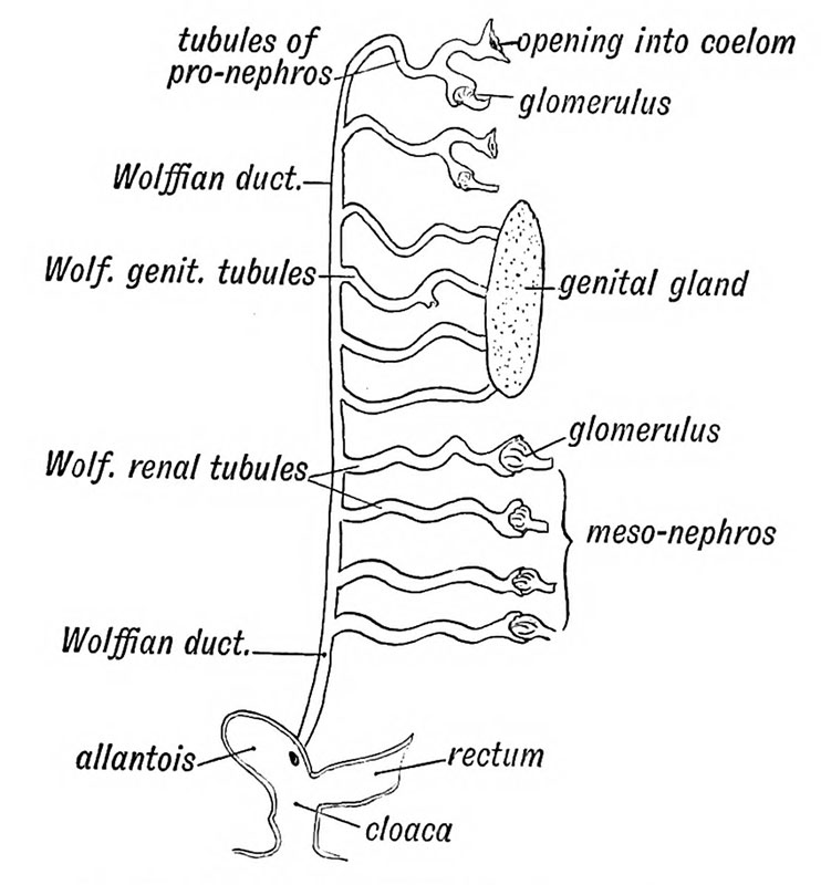 | 742 × 800 (78 KB) | Z8600021 (talk | contribs) |
You cannot overwrite this file.
File usage
The following 4 pages use this file: