File:Keith1902 fig055.jpg: Difference between revisions
No edit summary |
mNo edit summary |
||
| (2 intermediate revisions by the same user not shown) | |||
| Line 1: | Line 1: | ||
==Fig. 55 Diagram of a Developing Hair== | |||
Hairs begin to develop in the 5th month. Morphologically a hair may be regarded as a dermal papilla, which has become sunk in the subcutaneous tissue, and capped by a process of epidermis. Hairs appear to have been primarily touch organs and are modifications of the touch bodies found in the skin of Reptilia (Gegenbaur). These touch bodies are composed of epithelial cells, having the same shape and arrangement as those which form the taste buds round the circumvallate papillae of the human tongue. The cells which cap the hair papilla evidently represent the primary sensory cells of the touch bodies ; they are situated in line, and continuous with the basal or germinal layer of the skin (Fig. 55). They produce the cells of the medulla of the hair which bursts through the epidermis (Fig. 55). The primary function of the hairs as touch organs is seen in the vibrissae round the mouths of carnivora, but the hair of man no longer is subservient to the sense of touch. | |||
{{Keith1902_6_figures}} | |||
{{Human embryology morphology 1902 footer}} | |||
[[Category:Hair]] | |||
Latest revision as of 07:49, 23 January 2014
Fig. 55 Diagram of a Developing Hair
Hairs begin to develop in the 5th month. Morphologically a hair may be regarded as a dermal papilla, which has become sunk in the subcutaneous tissue, and capped by a process of epidermis. Hairs appear to have been primarily touch organs and are modifications of the touch bodies found in the skin of Reptilia (Gegenbaur). These touch bodies are composed of epithelial cells, having the same shape and arrangement as those which form the taste buds round the circumvallate papillae of the human tongue. The cells which cap the hair papilla evidently represent the primary sensory cells of the touch bodies ; they are situated in line, and continuous with the basal or germinal layer of the skin (Fig. 55). They produce the cells of the medulla of the hair which bursts through the epidermis (Fig. 55). The primary function of the hairs as touch organs is seen in the vibrissae round the mouths of carnivora, but the hair of man no longer is subservient to the sense of touch.
| Historic Disclaimer - information about historic embryology pages |
|---|
 |
- The Skin and its Appendages: Fig. 51. Skin strata first month | Fig. 52. Skin strata second month | Fig. 53. Skin strata sixth month onwards | Fig. 54. Common dermal papillae patterns on the finger tips | Fig. 55. Developing Hair | Fig. 56. Diagrammatic Section across a Nail | Fig. 57. Stages in Mamma development | Fig. 58. Section of the Breast | Figures
| Historic Disclaimer - information about historic embryology pages |
|---|
|
Human Embryology and Morphology (1902): Development or the Face | The Nasal Cavities and Olfactory Structures | Development of the Pharynx and Neck | Development of the Organ of Hearing | Development and Morphology of the Teeth | The Skin and its Appendages | The Development of the Ovum of the Foetus from the Ovum of the Mother | The Manner in which a Connection is Established between the Foetus and Uterus | The Uro-genital System | Formation of the Pubo-femoral Region, Pelvic Floor and Fascia | The Spinal Column and Back | The Segmentation of the Body | The Cranium | Development of the Structures concerned in the Sense of Sight | The Brain and Spinal Cord | Development of the Circulatory System | The Respiratory System | The Organs of Digestion | The Body Wall, Ribs, and Sternum | The Limbs | Figures | Embryology History
Reference
Keith A. Human Embryology and Morphology. (1902) London: Edward Arnold.
Cite this page: Hill, M.A. (2024, April 20) Embryology Keith1902 fig055.jpg. Retrieved from https://embryology.med.unsw.edu.au/embryology/index.php/File:Keith1902_fig055.jpg
- © Dr Mark Hill 2024, UNSW Embryology ISBN: 978 0 7334 2609 4 - UNSW CRICOS Provider Code No. 00098G
File history
Click on a date/time to view the file as it appeared at that time.
| Date/Time | Thumbnail | Dimensions | User | Comment | |
|---|---|---|---|---|---|
| current | 09:14, 7 January 2014 | 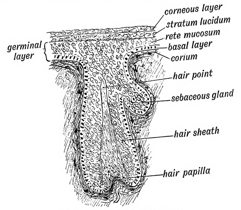 | 800 × 699 (136 KB) | Z8600021 (talk | contribs) |
You cannot overwrite this file.
File usage
The following 2 pages use this file: