File:Keith1902 fig040.jpg

Original file (1,000 × 524 pixels, file size: 85 KB, MIME type: image/jpeg)
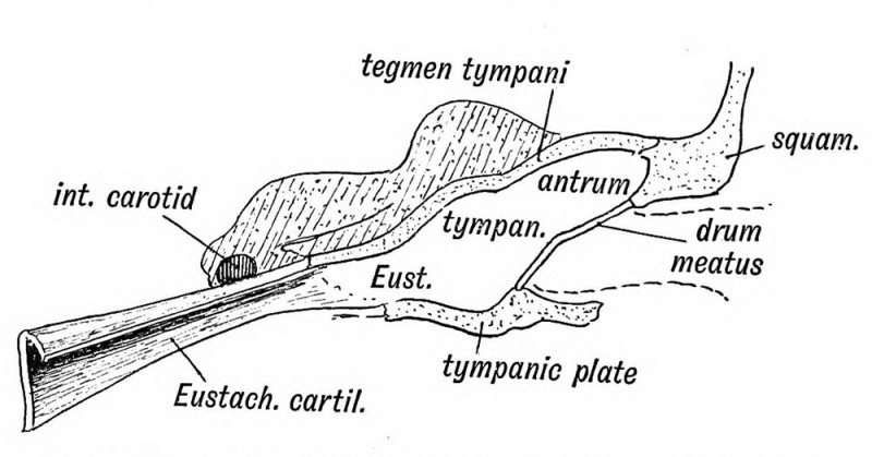
Fig. 40. Showing the Cavities derived from the Inner Recess of the First Cleft
The Eustachian tube is derived from the first inner cleft recess of the pharynx (Fig. 39) and retains through life the ciliated epithelial lining of the primitive pharynx. It is 1.45 inches long. Its inner two thirds is bounded behind by a triangular plate of cartilage, which is attached at its inner or pharyngeal end to the internal pterygoid plate, a derivative, with the incus, of the pterygo-palatine bar (Fig. 10 C). The cartilage is developed in the walls of the 1st visceral cleft in the 4th month of foetal life. The tympanic plate grows inwards and forms the floor of its outer third (Fig. 40), while the periotic capsule (petro-mastoid) which is developed above and behind the 1st cleft, grows over and forms the roof of its outer third. The part of the petro-mastoid which grows over it, is the tegmen tympani ; it also forms the roof of the tympanum and antrum of the mastoid. It forms the roof of all the cavities derived from the inner recess of the first cleft (Fig. 40). The anterior edge of the tegmen tympani appears in the Glaserian fissure. The tensor tympani and tensor palati are developed on the mandibular side of the first cleft and are supplied from the nerve of the mandibular process through the otic ganglion.
- Development of the Organ of Hearing: Fig. 35. Cephalic region of an embryo, showing the origin of the Auditory System | Fig. 36 A. Adult External Auditory Meatus | Fig. 36 B. External Auditory Meatus at Birth | Fig. 37. Tubercles round the First Visceral Cleft to form the External Ear | Fig. 38. Part of the Adult Ear formed by each Tubercle | Fig. 39. Auditory Organs 6th week human fetus | Fig. 40. Cavities from the Inner Recess of the First Cleft | Fig. 41. The temporal bone at birth | Fig. 42. Walls of the Antrum | Fig. 43. Outer aspect of the Petro-mastoid at birth | Fig. 44. Membranous Labyrinth | Fig. 45. The Otocyst in an Embryo of five weeks | Fig. 46. Nerve Structures Sense of Hearing | Figures
| Historic Disclaimer - information about historic embryology pages |
|---|
 |
Human Embryology and Morphology (1902): Development or the Face | The Nasal Cavities and Olfactory Structures | Development of the Pharynx and Neck | Development of the Organ of Hearing | Development and Morphology of the Teeth | The Skin and its Appendages | The Development of the Ovum of the Foetus from the Ovum of the Mother | The Manner in which a Connection is Established between the Foetus and Uterus | The Uro-genital System | Formation of the Pubo-femoral Region, Pelvic Floor and Fascia | The Spinal Column and Back | The Segmentation of the Body | The Cranium | Development of the Structures concerned in the Sense of Sight | The Brain and Spinal Cord | Development of the Circulatory System | The Respiratory System | The Organs of Digestion | The Body Wall, Ribs, and Sternum | The Limbs | Figures | Embryology History
Reference
Keith A. Human Embryology and Morphology. (1902) London: Edward Arnold.
Cite this page: Hill, M.A. (2024, April 19) Embryology Keith1902 fig040.jpg. Retrieved from https://embryology.med.unsw.edu.au/embryology/index.php/File:Keith1902_fig040.jpg
- © Dr Mark Hill 2024, UNSW Embryology ISBN: 978 0 7334 2609 4 - UNSW CRICOS Provider Code No. 00098G
File history
Click on a date/time to view the file as it appeared at that time.
| Date/Time | Thumbnail | Dimensions | User | Comment | |
|---|---|---|---|---|---|
| current | 02:38, 30 December 2013 | | 1,000 × 524 (85 KB) | Z8600021 (talk | contribs) | ==Fig. 40 Showing the Cavities derived from the Inner Recess of the First Cleft== {{Keith1902_4_figures}} {{Human embryology morphology 1902 footer}} |
You cannot overwrite this file.
File usage
The following 4 pages use this file: