File:Keith1902 fig039.jpg: Difference between revisions
mNo edit summary |
mNo edit summary |
||
| (3 intermediate revisions by the same user not shown) | |||
| Line 3: | Line 3: | ||
(After Siobenmann.) | (After Siobenmann.) | ||
As may be seen from Fig. 39, the tympanum can scarcely be said to exist at the sixth week of foetal life. The inner cleft recess ends in the jelly-like tissue containing the cartilaginous bases of the malleus and incus. It is directed outwards and backwards between the periotic capsule to its posterior and inner side and the external cleft depression (meatus) and developing squamosal to its outer (Fig. 39). As the internal recess extends and widens outwards and backwards, the gelatinous tissue is absorbed, so that the malleus and incus and developing stapes, with the chorda | As may be seen from [[:File:Keith1902 fig039.jpg|Fig. 39]], the tympanum can scarcely be said to exist at the sixth week of foetal life. The inner cleft recess ends in the jelly-like tissue containing the cartilaginous bases of the {{malleus}} and {{incus}}. It is directed outwards and backwards between the periotic capsule to its posterior and inner side and the external cleft depression (meatus) and developing squamosal to its outer ([[:File:Keith1902 fig039.jpg|Fig. 39]]). As the internal recess extends and widens outwards and backwards, the gelatinous tissue is absorbed, so that the malleus and incus and developing {{stapes}}, with the chorda tympani, become surrounded by the hypoblasts lining of the inner cleft recess and appear to be situated within the cavity thus formed — the tympanum. The tympanic plate forms its floor, the membrana tympani and squamosal its outer wall, while the petro-mastoid forms its inner wall and roof ([[:File:Keith1902 fig040.jpg|Fig. 40]]). That part of the tympanum which lies above the level of the membrana tympani is named the attic, and contains the head of the malleus and body of the incus ([[:File:Keith1902 fig036a.jpg|Fig. 36a]]). | ||
| Line 9: | Line 9: | ||
{{Human embryology morphology 1902 footer}} | {{Human embryology morphology 1902 footer}} | ||
[[Category:Human]][[Category:Week 6]] | |||
Latest revision as of 19:35, 17 April 2018
Fig. 39 Showing the condition of the Auditory Organs in a 6th week human foetus
(After Siobenmann.)
As may be seen from Fig. 39, the tympanum can scarcely be said to exist at the sixth week of foetal life. The inner cleft recess ends in the jelly-like tissue containing the cartilaginous bases of the malleus and incus. It is directed outwards and backwards between the periotic capsule to its posterior and inner side and the external cleft depression (meatus) and developing squamosal to its outer (Fig. 39). As the internal recess extends and widens outwards and backwards, the gelatinous tissue is absorbed, so that the malleus and incus and developing stapes, with the chorda tympani, become surrounded by the hypoblasts lining of the inner cleft recess and appear to be situated within the cavity thus formed — the tympanum. The tympanic plate forms its floor, the membrana tympani and squamosal its outer wall, while the petro-mastoid forms its inner wall and roof (Fig. 40). That part of the tympanum which lies above the level of the membrana tympani is named the attic, and contains the head of the malleus and body of the incus (Fig. 36a).
- Development of the Organ of Hearing: Fig. 35. Cephalic region of an embryo, showing the origin of the Auditory System | Fig. 36 A. Adult External Auditory Meatus | Fig. 36 B. External Auditory Meatus at Birth | Fig. 37. Tubercles round the First Visceral Cleft to form the External Ear | Fig. 38. Part of the Adult Ear formed by each Tubercle | Fig. 39. Auditory Organs 6th week human fetus | Fig. 40. Cavities from the Inner Recess of the First Cleft | Fig. 41. The temporal bone at birth | Fig. 42. Walls of the Antrum | Fig. 43. Outer aspect of the Petro-mastoid at birth | Fig. 44. Membranous Labyrinth | Fig. 45. The Otocyst in an Embryo of five weeks | Fig. 46. Nerve Structures Sense of Hearing | Figures
| Historic Disclaimer - information about historic embryology pages |
|---|
 |
Human Embryology and Morphology (1902): Development or the Face | The Nasal Cavities and Olfactory Structures | Development of the Pharynx and Neck | Development of the Organ of Hearing | Development and Morphology of the Teeth | The Skin and its Appendages | The Development of the Ovum of the Foetus from the Ovum of the Mother | The Manner in which a Connection is Established between the Foetus and Uterus | The Uro-genital System | Formation of the Pubo-femoral Region, Pelvic Floor and Fascia | The Spinal Column and Back | The Segmentation of the Body | The Cranium | Development of the Structures concerned in the Sense of Sight | The Brain and Spinal Cord | Development of the Circulatory System | The Respiratory System | The Organs of Digestion | The Body Wall, Ribs, and Sternum | The Limbs | Figures | Embryology History
Reference
Keith A. Human Embryology and Morphology. (1902) London: Edward Arnold.
Cite this page: Hill, M.A. (2024, April 24) Embryology Keith1902 fig039.jpg. Retrieved from https://embryology.med.unsw.edu.au/embryology/index.php/File:Keith1902_fig039.jpg
- © Dr Mark Hill 2024, UNSW Embryology ISBN: 978 0 7334 2609 4 - UNSW CRICOS Provider Code No. 00098G
File history
Click on a date/time to view the file as it appeared at that time.
| Date/Time | Thumbnail | Dimensions | User | Comment | |
|---|---|---|---|---|---|
| current | 02:29, 30 December 2013 | 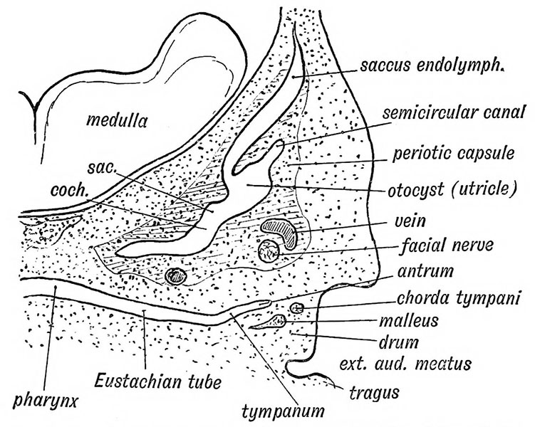 | 751 × 600 (114 KB) | Z8600021 (talk | contribs) | ==Fig. 39 Showing the condition of the Auditory Organs in a 6th week human foetus== (After Siobenmann.) {{Keith1902_4_figures}} {{Human embryology morphology 1902 footer}} |
You cannot overwrite this file.
File usage
The following 4 pages use this file: