File:Keibel Mall 315.jpg
From Embryology
No higher resolution available.
Keibel_Mall_315.jpg (736 × 433 pixels, file size: 84 KB, MIME type: image/jpeg)
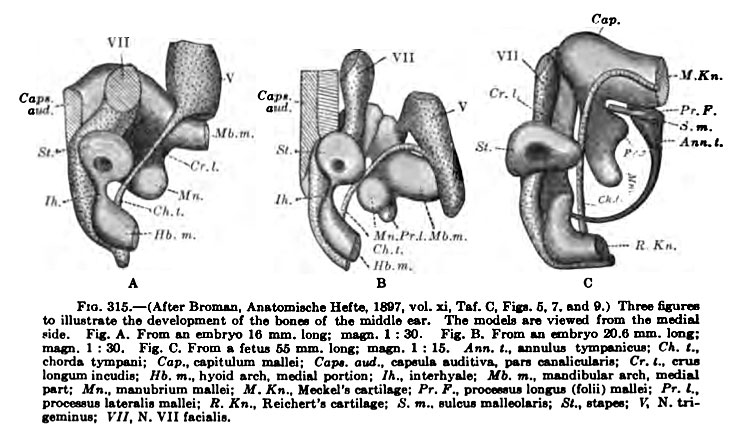
Fig. 313 Human Embryo Skull Middle Ear
Three figures to illustrate the development of the bones of the middle ear. The models are viewed from the medial side.
- Fig. A - From an embryo 16 mm long
- Fig. B - From an embryo 20.6 mm long
- Fig. C - From an embryo 55 mm long
- Skull Images: 308 | 309 | 310 | 311 | 312 | 313 | 314 | 315 | 316 | 317 | 318 | 319 | 320 | 321 | 322 | 323 | 324 | Text | Skull Development
- KM Figure Links: The Germ Cells | Segmentation | First Primitive Segment | Gastrulation | External Form | Placenta | Axial Skeleton | Limb Skeleton | Skull | Muscular System
| Historic Disclaimer - information about historic embryology pages |
|---|
 |
Glossary Links
- Glossary: A | B | C | D | E | F | G | H | I | J | K | L | M | N | O | P | Q | R | S | T | U | V | W | X | Y | Z | Numbers | Symbols | Term Link
Cite this page: Hill, M.A. (2024, April 18) Embryology Keibel Mall 315.jpg. Retrieved from https://embryology.med.unsw.edu.au/embryology/index.php/File:Keibel_Mall_315.jpg
- © Dr Mark Hill 2024, UNSW Embryology ISBN: 978 0 7334 2609 4 - UNSW CRICOS Provider Code No. 00098G
File history
Click on a date/time to view the file as it appeared at that time.
| Date/Time | Thumbnail | Dimensions | User | Comment | |
|---|---|---|---|---|---|
| current | 09:32, 27 August 2012 | | 736 × 433 (84 KB) | Z8600021 (talk | contribs) | ==Fig. 313 Human Embryo Skull== Visceral skeleton of the labyrinth of a human fetus at the end of the third month (8 cm long). (After Hertwig's model, from Kollman Handatlas, 1907, Fig. 266) {{KM Skull}} {{Keibel_Mall Images}} Category:Human [[C |
You cannot overwrite this file.
File usage
The following 4 pages use this file: