File:Keibel Mall 308.jpg
Keibel_Mall_308.jpg (677 × 578 pixels, file size: 43 KB, MIME type: image/jpeg)
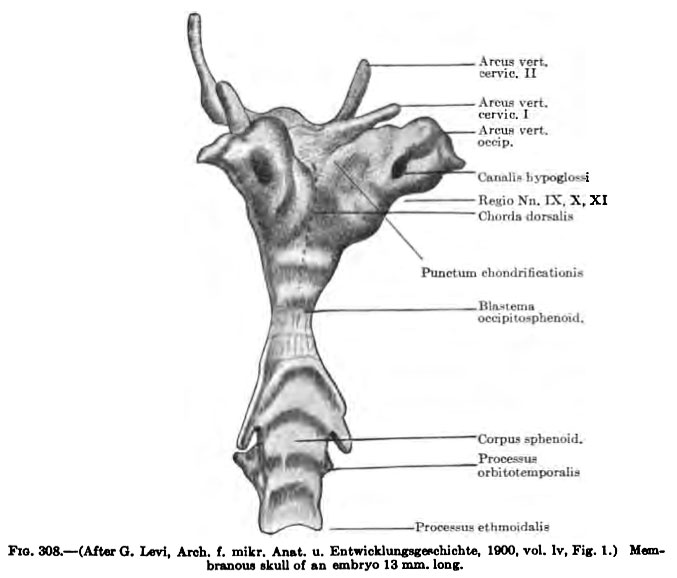
Fig. 308 Human Embryo Skull
Membranous skull of an embryo 13 mm long.
The anterior and posterior lateral processes of the occipital plate become united lateral to the hypoglossal nerve, so that the hypoglossal foramen is completed and the membranous pars lateralis of the occipital is formed. This pars lateralis is continued into the membranous vault of the skull, the origin of which is described below. The condensed tissue of the post-hypophyseal region increases in amount and extends about the hypophyseal pocket into the region apical from this, thus completing the anlage of the body of the sphenoid {Fig. 308). This gives rise to orbitotemporal and ethmoidal processes. The orbitotemporal process is first marked by a mass of dense mesenchyme which extends ventrolaterally toward the ectoderm caudal to the optic cup. It is connected with dense tissue which surrounds the anlage of the orbit and with the anlages of the membranous fioor and vault of the skull. In it are developed the orbital and temporal wings of the sphenoid, the origin of which will be described in connection with the chondroeranium. The ethmoidal process extends anteriorly in the median line from the anlage of the body of the sphenoid into the region between the nasal fosste. It forms the anlage of the nasal septum and gives rise to parts of the membranous floor of the cranial cavity and the roof of the mouth (Fig. 310).
- Skull Images: 308 | 309 | 310 | 311 | 312 | 313 | 314 | 315 | 316 | 317 | 318 | 319 | 320 | 321 | 322 | 323 | 324 | Text | Skull Development
- KM Figure Links: The Germ Cells | Segmentation | First Primitive Segment | Gastrulation | External Form | Placenta | Axial Skeleton | Limb Skeleton | Skull | Muscular System
| Historic Disclaimer - information about historic embryology pages |
|---|
 |
Glossary Links
- Glossary: A | B | C | D | E | F | G | H | I | J | K | L | M | N | O | P | Q | R | S | T | U | V | W | X | Y | Z | Numbers | Symbols | Term Link
Cite this page: Hill, M.A. (2024, April 16) Embryology Keibel Mall 308.jpg. Retrieved from https://embryology.med.unsw.edu.au/embryology/index.php/File:Keibel_Mall_308.jpg
- © Dr Mark Hill 2024, UNSW Embryology ISBN: 978 0 7334 2609 4 - UNSW CRICOS Provider Code No. 00098G
File history
Click on a date/time to view the file as it appeared at that time.
| Date/Time | Thumbnail | Dimensions | User | Comment | |
|---|---|---|---|---|---|
| current | 08:46, 27 August 2012 | | 677 × 578 (43 KB) | Z8600021 (talk | contribs) |
You cannot overwrite this file.
File usage
The following 4 pages use this file: