File:Keibel Mall 2 656.jpg: Difference between revisions
(==Fig. 656. == {{Human Embryology Manual 2 19}} {{Keibel_Mall 2 Images}}) |
mNo edit summary |
||
| (2 intermediate revisions by the same user not shown) | |||
| Line 1: | Line 1: | ||
==Fig. 656. == | ==Fig. 656. The various parts of the chorda gubernaculi in a transverse section of the urogenital fold and body wall of a male embryo of 26 mm greatest length== | ||
The chorda gubernaculi in the broader sense consists of: | |||
# the lig. testis (''pars mesorchica'') | |||
# a mesenchymatous cord in the mesonephric fold and its plica inguinalis (''pars mesonephridica'') | |||
# a mesenchymatous cord in the crista inguinalis and between the abdominal muscles (''pars intermuseularis'') | |||
# the lig. scroti (''pars scrotalis''). | |||
| Line 8: | Line 14: | ||
{{Keibel_Mall 2 Images}} | {{Keibel_Mall 2 Images}} | ||
[[Category:Human]] [[Category:Genital]] [[Category:Male]] | |||
Latest revision as of 12:18, 19 February 2014
Fig. 656. The various parts of the chorda gubernaculi in a transverse section of the urogenital fold and body wall of a male embryo of 26 mm greatest length
The chorda gubernaculi in the broader sense consists of:
- the lig. testis (pars mesorchica)
- a mesenchymatous cord in the mesonephric fold and its plica inguinalis (pars mesonephridica)
- a mesenchymatous cord in the crista inguinalis and between the abdominal muscles (pars intermuseularis)
- the lig. scroti (pars scrotalis).
| Embryology - 20 Apr 2024 |
|---|
| Google Translate - select your language from the list shown below (this will open a new external page) |
|
العربية | català | 中文 | 中國傳統的 | français | Deutsche | עִברִית | हिंदी | bahasa Indonesia | italiano | 日本語 | 한국어 | မြန်မာ | Pilipino | Polskie | português | ਪੰਜਾਬੀ ਦੇ | Română | русский | Español | Swahili | Svensk | ไทย | Türkçe | اردو | ייִדיש | Tiếng Việt These external translations are automated and may not be accurate. (More? About Translations) |
Felix W. The development of the urinogenital organs. In Keibel F. and Mall FP. Manual of Human Embryology II. (1912) J. B. Lippincott Company, Philadelphia. pp 752-979.
| Historic Disclaimer - information about historic embryology pages |
|---|
 |
Cite this page: Hill, M.A. (2024, April 20) Embryology Keibel Mall 2 656.jpg. Retrieved from https://embryology.med.unsw.edu.au/embryology/index.php/File:Keibel_Mall_2_656.jpg
- © Dr Mark Hill 2024, UNSW Embryology ISBN: 978 0 7334 2609 4 - UNSW CRICOS Provider Code No. 00098G
Manual of Human Embryology II: Nervous System | Chromaffin Organs and Suprarenal Bodies | Sense-Organs | Digestive Tract and Respiration | Vascular System | Urinogenital Organs | Figures 2 | Manual of Human Embryology 1 | Figures 1 | Manual of Human Embryology 2 | Figures 2 | Franz Keibel | Franklin Mall | Embryology History
Cite this page: Hill, M.A. (2024, April 20) Embryology Keibel Mall 2 656.jpg. Retrieved from https://embryology.med.unsw.edu.au/embryology/index.php/File:Keibel_Mall_2_656.jpg
- © Dr Mark Hill 2024, UNSW Embryology ISBN: 978 0 7334 2609 4 - UNSW CRICOS Provider Code No. 00098G
File history
Click on a date/time to view the file as it appeared at that time.
| Date/Time | Thumbnail | Dimensions | User | Comment | |
|---|---|---|---|---|---|
| current | 21:35, 9 February 2014 | 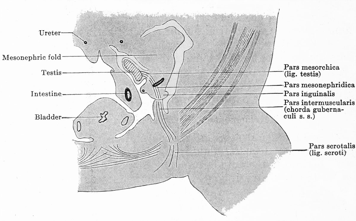 | 1,200 × 746 (152 KB) | Z8600021 (talk | contribs) | ==Fig. 656. == {{Human Embryology Manual 2 19}} {{Keibel_Mall 2 Images}} |
You cannot overwrite this file.
File usage
The following 2 pages use this file: