File:Keibel Mall 2 609.jpg

Original file (1,200 × 753 pixels, file size: 130 KB, MIME type: image/jpeg)
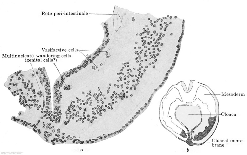
Fig. 609 a and b. Part of a transverse section through the embryo 2.5 mm greatest length 13-14 pairs of primitive segments
Embryo Pfannenstiel III from the collection of the late Professor Pfannenstiel, Kiel.)
The section passes through the cloacal membrane and its exact position may be seen from the adjacent figure b. The mesoderm is still quite loosely aggregated. Between it and the entoderm there is at the upper edge of the figure a fully-formed portion of the rete peri-intestinale, and further down there are cell chains and masses (to the left of the cloaca) from which new networks of the rete are forming. The section shows at two places, to the right and left of the cloacal membrane, large cells partly multinucleate, partly filled with yolk granules and partly free from them, and lying free between the mesoderm and endoderm. These cells may be termed wandering cells and hypothetically may be interpreted as primary genital cells.
File history
Click on a date/time to view the file as it appeared at that time.
| Date/Time | Thumbnail | Dimensions | User | Comment | |
|---|---|---|---|---|---|
| current | 07:22, 20 February 2014 | | 1,200 × 753 (130 KB) | Z8600021 (talk | contribs) | ==Fig. 609 a and b. Part of a transverse section through the embryo Pfannenstiel III, 2.5 mm greatest length and with 13-14 pairs of primitive segments== (From the collection of the late Professor Pfannenstiel, Kiel.) The section passes through the ... |
You cannot overwrite this file.
File usage
The following 2 pages use this file: