File:Keibel Mall 2 576.jpg: Difference between revisions
(Z8600021 uploaded a new version of File:Keibel Mall 2 576.jpg) |
mNo edit summary |
||
| Line 1: | Line 1: | ||
==Fig. 576. Reconstruction of the lower end of the body of a human embryo of 5.3 mm greatest length== | ==Fig. 576. Reconstruction of the lower end of the body of a human embryo of 5.3 mm greatest length== | ||
4.6 mm nape length, and with 36 pairs of primitive segments. (Embryo | 4.6 mm nape length, and with 36 pairs of primitive segments. (Embryo {{KE1420}}, from the collection of Professor Keibel, Freiburg i. Br.) | ||
The primary excretory duct has come to lie between the visceral and the parietal roots of the a. umbilicalis. At the level of its opening into the bladder it bends at a right angle and at the bend it pushes out the ureter bud stages of the development of the ureter. The hemispherical ureteric anlage is not exactly in the middle of the dorsal wall of the primary duct, but inclines more and more towards its medial surface, the degree of inclination varying in different embryos. When the anlage later elongates to form a canal it always lies at the middle of the dorsal surface of primary duct and, later on, even on its lateral surface, yet in an embryo of 7.8 mm. greatest length it still opened distinctly on the medial surface. | The primary excretory duct has come to lie between the visceral and the parietal roots of the a. umbilicalis. At the level of its opening into the bladder it bends at a right angle and at the bend it pushes out the ureter bud stages of the development of the ureter. The hemispherical ureteric anlage is not exactly in the middle of the dorsal wall of the primary duct, but inclines more and more towards its medial surface, the degree of inclination varying in different embryos. When the anlage later elongates to form a canal it always lies at the middle of the dorsal surface of primary duct and, later on, even on its lateral surface, yet in an embryo of 7.8 mm. greatest length it still opened distinctly on the medial surface. | ||
Latest revision as of 10:51, 13 November 2018
Fig. 576. Reconstruction of the lower end of the body of a human embryo of 5.3 mm greatest length
4.6 mm nape length, and with 36 pairs of primitive segments. (Embryo 1420, from the collection of Professor Keibel, Freiburg i. Br.)
The primary excretory duct has come to lie between the visceral and the parietal roots of the a. umbilicalis. At the level of its opening into the bladder it bends at a right angle and at the bend it pushes out the ureter bud stages of the development of the ureter. The hemispherical ureteric anlage is not exactly in the middle of the dorsal wall of the primary duct, but inclines more and more towards its medial surface, the degree of inclination varying in different embryos. When the anlage later elongates to form a canal it always lies at the middle of the dorsal surface of primary duct and, later on, even on its lateral surface, yet in an embryo of 7.8 mm. greatest length it still opened distinctly on the medial surface.
| Embryology - 16 Apr 2024 |
|---|
| Google Translate - select your language from the list shown below (this will open a new external page) |
|
العربية | català | 中文 | 中國傳統的 | français | Deutsche | עִברִית | हिंदी | bahasa Indonesia | italiano | 日本語 | 한국어 | မြန်မာ | Pilipino | Polskie | português | ਪੰਜਾਬੀ ਦੇ | Română | русский | Español | Swahili | Svensk | ไทย | Türkçe | اردو | ייִדיש | Tiếng Việt These external translations are automated and may not be accurate. (More? About Translations) |
Felix W. The development of the urinogenital organs. In Keibel F. and Mall FP. Manual of Human Embryology II. (1912) J. B. Lippincott Company, Philadelphia. pp 752-979.
| Historic Disclaimer - information about historic embryology pages |
|---|
 |
Cite this page: Hill, M.A. (2024, April 16) Embryology Keibel Mall 2 576.jpg. Retrieved from https://embryology.med.unsw.edu.au/embryology/index.php/File:Keibel_Mall_2_576.jpg
- © Dr Mark Hill 2024, UNSW Embryology ISBN: 978 0 7334 2609 4 - UNSW CRICOS Provider Code No. 00098G
File history
Click on a date/time to view the file as it appeared at that time.
| Date/Time | Thumbnail | Dimensions | User | Comment | |
|---|---|---|---|---|---|
| current | 10:42, 13 November 2018 |  | 1,280 × 767 (154 KB) | Z8600021 (talk | contribs) | |
| 10:36, 13 November 2018 | 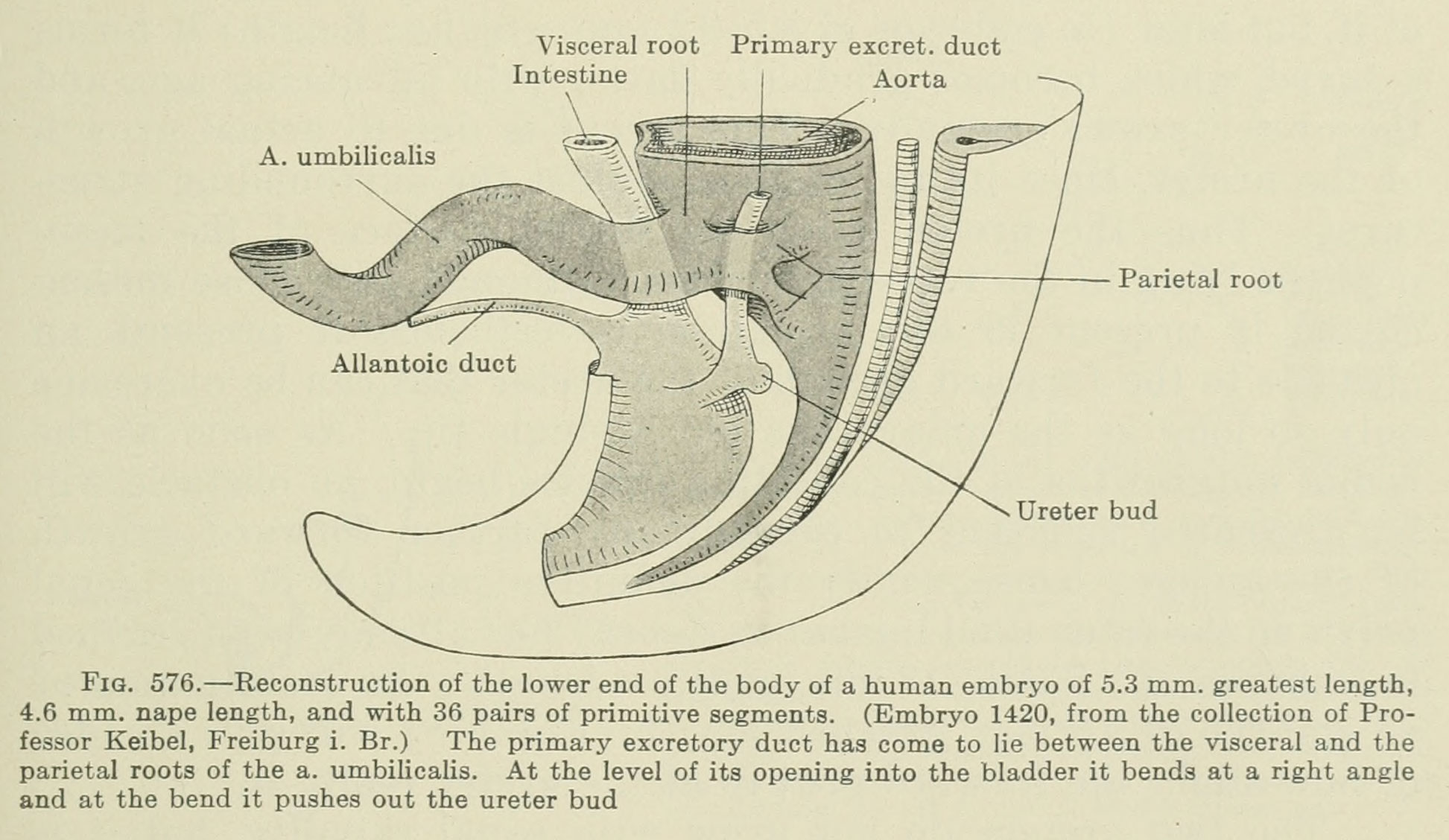 | 1,945 × 1,128 (258 KB) | Z8600021 (talk | contribs) |
You cannot overwrite this file.
File usage
The following 2 pages use this file: