File:Keibel Mall 2 552.jpg: Difference between revisions
(Z8600021 uploaded a new version of File:Keibel Mall 2 552.jpg) |
mNo edit summary |
||
| Line 3: | Line 3: | ||
(Embryo Ma. 2, from the collection of Professor Hochstetter, Berlin. The model was prepared by my students Massard and Chome.) | (Embryo Ma. 2, from the collection of Professor Hochstetter, Berlin. The model was prepared by my students Massard and Chome.) | ||
The visceral layer of peritoneum is shown cut and one sees the superior recess of the omental sack. The urogenital fold is divided as far as its upper and lower ends into the mesonephric and genital folds. The mesonephric fold is bayonet-shaped and an upper sagittal, a horizontal and a lower sagittal portion, and a first and second bend may be distinguished. At the first bend the mesonephric fold is connected with the anterior abdominal wall by the inguinal fold. Between the aa. umbilicales, which still run horizontally, is the bladder plate. | The visceral layer of peritoneum is shown cut and one sees the superior recess of the omental sack. The urogenital fold is divided as far as its upper and lower ends into the mesonephric and genital folds. The mesonephric fold is bayonet-shaped and an upper sagittal, a horizontal and a lower sagittal portion, and a first and second bend may be distinguished. At the first bend the mesonephric fold is connected with the anterior abdominal wall by the inguinal fold. Between the aa. umbilicales, which still run horizontally, is the {{bladder}} plate. | ||
{{Human Embryology Manual 2 19}} | |||
[[Category:Renal]][[Category:Genital]] | |||
Latest revision as of 07:41, 16 November 2018
Fig. 552. Model of the posterior abdominal wall of a human embryo of 19.4 mm greatest length
(Embryo Ma. 2, from the collection of Professor Hochstetter, Berlin. The model was prepared by my students Massard and Chome.)
The visceral layer of peritoneum is shown cut and one sees the superior recess of the omental sack. The urogenital fold is divided as far as its upper and lower ends into the mesonephric and genital folds. The mesonephric fold is bayonet-shaped and an upper sagittal, a horizontal and a lower sagittal portion, and a first and second bend may be distinguished. At the first bend the mesonephric fold is connected with the anterior abdominal wall by the inguinal fold. Between the aa. umbilicales, which still run horizontally, is the bladder plate.
| Embryology - 18 Apr 2024 |
|---|
| Google Translate - select your language from the list shown below (this will open a new external page) |
|
العربية | català | 中文 | 中國傳統的 | français | Deutsche | עִברִית | हिंदी | bahasa Indonesia | italiano | 日本語 | 한국어 | မြန်မာ | Pilipino | Polskie | português | ਪੰਜਾਬੀ ਦੇ | Română | русский | Español | Swahili | Svensk | ไทย | Türkçe | اردو | ייִדיש | Tiếng Việt These external translations are automated and may not be accurate. (More? About Translations) |
Felix W. The development of the urinogenital organs. In Keibel F. and Mall FP. Manual of Human Embryology II. (1912) J. B. Lippincott Company, Philadelphia. pp 752-979.
| Historic Disclaimer - information about historic embryology pages |
|---|
 |
Cite this page: Hill, M.A. (2024, April 18) Embryology Keibel Mall 2 552.jpg. Retrieved from https://embryology.med.unsw.edu.au/embryology/index.php/File:Keibel_Mall_2_552.jpg
- © Dr Mark Hill 2024, UNSW Embryology ISBN: 978 0 7334 2609 4 - UNSW CRICOS Provider Code No. 00098G
File history
Click on a date/time to view the file as it appeared at that time.
| Date/Time | Thumbnail | Dimensions | User | Comment | |
|---|---|---|---|---|---|
| current | 07:12, 15 November 2018 | 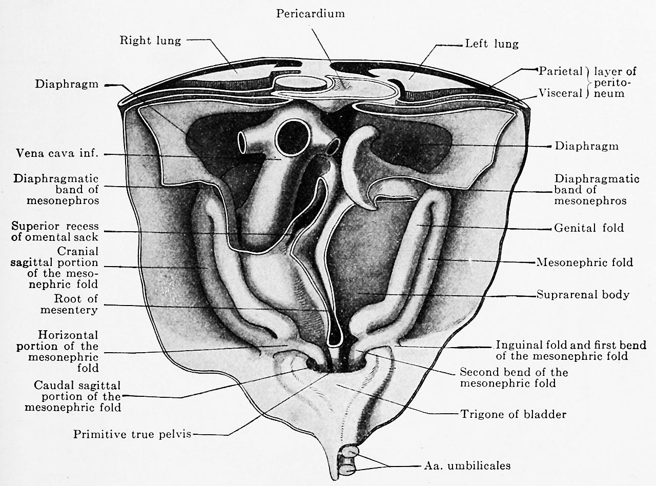 | 1,280 × 948 (257 KB) | Z8600021 (talk | contribs) | |
| 07:07, 15 November 2018 | 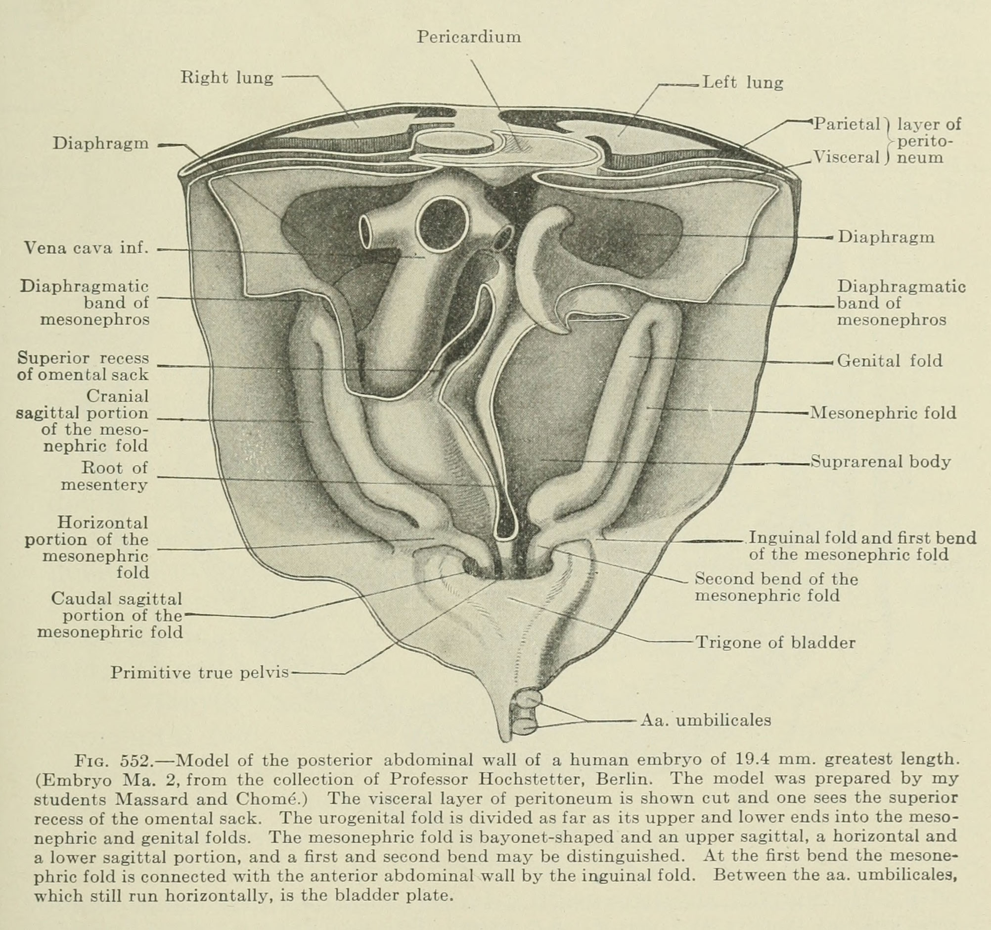 | 2,004 × 1,879 (588 KB) | Z8600021 (talk | contribs) |
You cannot overwrite this file.
File usage
The following 2 pages use this file: