File:Keibel Mall 2 539.jpg: Difference between revisions
Original file (1,280 × 1,414 pixels, file size: 185 KB, MIME type: image/jpeg)
mNo edit summary |
mNo edit summary |
||
| (One intermediate revision by the same user not shown) | |||
| Line 9: | Line 9: | ||
{{Human Embryology Manual 2 19}} | {{Human Embryology Manual 2 19}} | ||
[[Category:Artery]][[Category:Cardiovascular]][[Category:Yolk Sac]] | [[Category:Artery]][[Category:Cardiovascular]][[Category:Yolk Sac]] | ||
[[Category:Carnegie Stage 10]] | |||
Latest revision as of 18:25, 14 November 2018
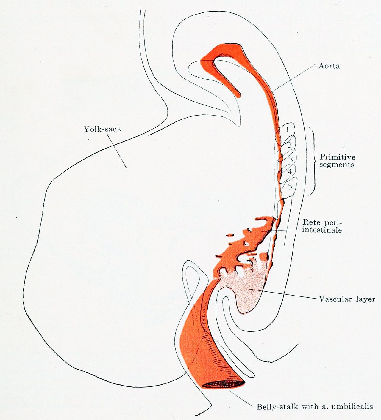
Fig. 539. Reconstruction of the arterial system of an embryo of 1.38 mm greatest length
with 5-6 pairs of primitive segments. (Embryo Pfannenstiel-Kromer, from the collection of Professor Pfannenstiel.)
The aorta is a continuous vessel with completely closed walls from the level of the second primitive segment forward, from there backwards it is in anlage. On the posterior part of the yolk-sack and on the end-gut is the anlage of the rete peri-intestinale. The a. umbilicalis arises from this rete.
| Embryology - 18 Apr 2024 |
|---|
| Google Translate - select your language from the list shown below (this will open a new external page) |
|
العربية | català | 中文 | 中國傳統的 | français | Deutsche | עִברִית | हिंदी | bahasa Indonesia | italiano | 日本語 | 한국어 | မြန်မာ | Pilipino | Polskie | português | ਪੰਜਾਬੀ ਦੇ | Română | русский | Español | Swahili | Svensk | ไทย | Türkçe | اردو | ייִדיש | Tiếng Việt These external translations are automated and may not be accurate. (More? About Translations) |
Felix W. The development of the urinogenital organs. In Keibel F. and Mall FP. Manual of Human Embryology II. (1912) J. B. Lippincott Company, Philadelphia. pp 752-979.
| Historic Disclaimer - information about historic embryology pages |
|---|
 |
Cite this page: Hill, M.A. (2024, April 18) Embryology Keibel Mall 2 539.jpg. Retrieved from https://embryology.med.unsw.edu.au/embryology/index.php/File:Keibel_Mall_2_539.jpg
- © Dr Mark Hill 2024, UNSW Embryology ISBN: 978 0 7334 2609 4 - UNSW CRICOS Provider Code No. 00098G
File history
Click on a date/time to view the file as it appeared at that time.
| Date/Time | Thumbnail | Dimensions | User | Comment | |
|---|---|---|---|---|---|
| current | 17:58, 14 November 2018 | | 1,280 × 1,414 (185 KB) | Z8600021 (talk | contribs) | |
| 17:56, 14 November 2018 | 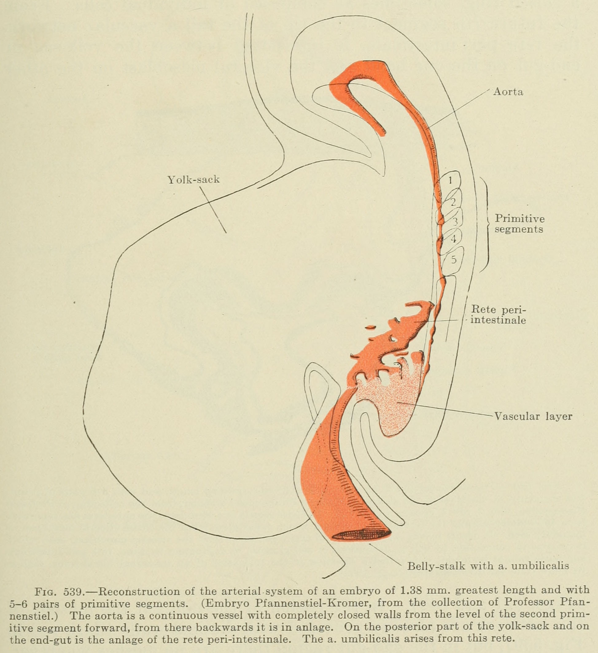 | 1,948 × 2,127 (383 KB) | Z8600021 (talk | contribs) | {{Human Embryology Manual 2 19}} Category:Renal |
You cannot overwrite this file.
File usage
The following 3 pages use this file: