File:Keibel Mall 2 526.jpg: Difference between revisions
mNo edit summary |
mNo edit summary |
||
| Line 1: | Line 1: | ||
==Fig. 526. Reconstruction of the right pronephric anlage of an embryo of 2.6 mm greatest length== | ==Fig. 526. Reconstruction of the right pronephric anlage of an embryo of 2.6 mm greatest length== | ||
With 13-14 pairs of primitive segments. (Embryo Pfannenstiel III, from the collection of Professor Pfannenstiel.) | With 13-14 pairs of primitive segments. (Embryo {{Pfannenstiel III}}, from the collection of Professor Pfannenstiel.) | ||
The contours of the embryo, the medullary canal and the intestine are outlined, the mesoderm (secondary primitive segment, segment stalk and lateral plate) is shaded, the pronephric anlage is black. The anlage consists of five parts, a rudimentary tubule in the 7th segment, two well-developed tubules in the 8th and 9th segments, a pronephric ridge in the 10th, 11th and 12th segments and another ridge in the 13th and 14th segments. Luminain the principal tubule and in the primitive segmentjstalks are left white. | The contours of the embryo, the medullary canal and the intestine are outlined, the {{mesoderm}} (secondary primitive segment, segment stalk and lateral plate) is shaded, the pronephric anlage is black. The anlage consists of five parts, a rudimentary tubule in the 7th segment, two well-developed tubules in the 8th and 9th segments, a pronephric ridge in the 10th, 11th and 12th segments and another ridge in the 13th and 14th segments. Luminain the principal tubule and in the primitive segmentjstalks are left white. | ||
{{Human Embryology Manual 2 19}} | {{Human Embryology Manual 2 19}} | ||
[[Category:Renal]] | [[Category:Renal]][[Category:Pfannenstiel III]] | ||
Latest revision as of 18:35, 23 June 2019
Fig. 526. Reconstruction of the right pronephric anlage of an embryo of 2.6 mm greatest length
With 13-14 pairs of primitive segments. (Embryo Pfannenstiel III, from the collection of Professor Pfannenstiel.)
The contours of the embryo, the medullary canal and the intestine are outlined, the mesoderm (secondary primitive segment, segment stalk and lateral plate) is shaded, the pronephric anlage is black. The anlage consists of five parts, a rudimentary tubule in the 7th segment, two well-developed tubules in the 8th and 9th segments, a pronephric ridge in the 10th, 11th and 12th segments and another ridge in the 13th and 14th segments. Luminain the principal tubule and in the primitive segmentjstalks are left white.
| Embryology - 23 Apr 2024 |
|---|
| Google Translate - select your language from the list shown below (this will open a new external page) |
|
العربية | català | 中文 | 中國傳統的 | français | Deutsche | עִברִית | हिंदी | bahasa Indonesia | italiano | 日本語 | 한국어 | မြန်မာ | Pilipino | Polskie | português | ਪੰਜਾਬੀ ਦੇ | Română | русский | Español | Swahili | Svensk | ไทย | Türkçe | اردو | ייִדיש | Tiếng Việt These external translations are automated and may not be accurate. (More? About Translations) |
Felix W. The development of the urinogenital organs. In Keibel F. and Mall FP. Manual of Human Embryology II. (1912) J. B. Lippincott Company, Philadelphia. pp 752-979.
| Historic Disclaimer - information about historic embryology pages |
|---|
 |
Cite this page: Hill, M.A. (2024, April 23) Embryology Keibel Mall 2 526.jpg. Retrieved from https://embryology.med.unsw.edu.au/embryology/index.php/File:Keibel_Mall_2_526.jpg
- © Dr Mark Hill 2024, UNSW Embryology ISBN: 978 0 7334 2609 4 - UNSW CRICOS Provider Code No. 00098G
File history
Click on a date/time to view the file as it appeared at that time.
| Date/Time | Thumbnail | Dimensions | User | Comment | |
|---|---|---|---|---|---|
| current | 16:49, 14 November 2018 | 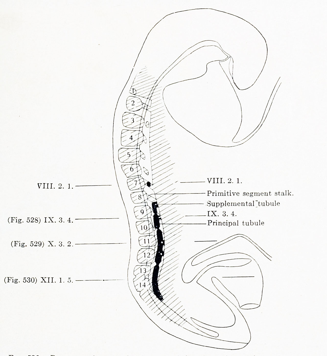 | 1,280 × 1,391 (186 KB) | Z8600021 (talk | contribs) | |
| 16:48, 14 November 2018 |  | 1,966 × 2,036 (492 KB) | Z8600021 (talk | contribs) |
You cannot overwrite this file.
File usage
The following 2 pages use this file: