File:Keibel Mall 2 302.jpg: Difference between revisions
mNo edit summary |
mNo edit summary |
||
| (One intermediate revision by the same user not shown) | |||
| Line 22: | Line 22: | ||
[[Category:Liver]] | [[Category:Liver]][[Category:Fetal]][[Category:Second Trimester]] | ||
Latest revision as of 16:30, 4 February 2019
Fig. 302. Dorsal views of the hepatic region
Fig. 302, dissection of a 5 months' embryo, 220 mm in length, X l.33 diam.
ap.cor. d., ap. cor. s., right and left coronary appendages; c. pi., pleural part of the pleuroperitoneal cavity; c. per., peritoneal part of the pleuroperitoneal cavity; d. ch., common bile-duct; dia., diaphragm; for. ep., foramen epiploicum; I. cor., coronary ligament; 1. hep, d., I. hep. «., right and left hepatic lobes; mes., mesentery; oes., oesophagus; om. m., lesser omentum; pl. v. c, plica venae cavae; r. s., superior lateral recess of the peritoneal cavity; s.tr. septum transversum; v.c, vena cava; ves. fel., gall-bladder; v. p., portal vein; v. um., umbilical vein.
In an embryo of 5 months (Fig. 302) the diaphragm has been completed in the way described in Chapter XIII. The oesophagus, not included in the preceding drawings, is seen passing through it. The thin lateral extensions of each coronary ligament {ligamenta triangularia) mark the position of the former appendages, and filling their dorsal concavity is the portion of the diaphragm which formed last, and which completes the separation of pleural and peritoneal cavities. The vena cava inferior now fills its plica, which has become broad. The lesser omentum is very thin except 10 They were probably included by Lieberkuhn (1876) among 1 the "villi" which occur where the omphalomesenteric veins enter the heart, and which were said to be so related to the developing liver that they contained the first blood vessels of that organ.
Fig. 300 4 mm embryo | Fig. 301 9.4 mm embryo | Fig. 302 5 months' embryo
| Historic Disclaimer - information about historic embryology pages |
|---|
 |
Reference
Grosser O. Lewis FT. and McMurrich JP. The Development of the Digestive Tract and of the Organs of Respiration. (1912) chapter 17, vol. 2, in Keibel F. and Mall FP. Manual of Human Embryology II. (1912) J. B. Lippincott Company, Philadelphia.
Cite this page: Hill, M.A. (2024, April 19) Embryology Keibel Mall 2 302.jpg. Retrieved from https://embryology.med.unsw.edu.au/embryology/index.php/File:Keibel_Mall_2_302.jpg
- © Dr Mark Hill 2024, UNSW Embryology ISBN: 978 0 7334 2609 4 - UNSW CRICOS Provider Code No. 00098G
File history
Click on a date/time to view the file as it appeared at that time.
| Date/Time | Thumbnail | Dimensions | User | Comment | |
|---|---|---|---|---|---|
| current | 16:13, 4 February 2019 | 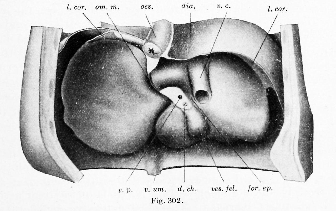 | 1,278 × 803 (149 KB) | Z8600021 (talk | contribs) |
You cannot overwrite this file.
File usage
The following 2 pages use this file: