File:Keibel Mall 2 301.jpg: Difference between revisions
mNo edit summary |
mNo edit summary |
||
| Line 5: | Line 5: | ||
ap.cor. d., ap. cor. s., right and left coronary appendages; c. pi., pleural part of the pleuroperitoneal cavity; c. per., peritoneal part of the pleuroperitoneal cavity; d. ch., common bile-duct; dia., diaphragm; for. ep., foramen epiploicum; I. cor., coronary ligament; 1. hep, d., I. hep. «., right and left hepatic lobes; mes., mesentery; oes., oesophagus; om. m., lesser omentum; pl. v. c, plica venae cavae; r. s., superior lateral recess of the peritoneal cavity; s.tr. septum transversum; v.c, vena cava; ves. fel., gall-bladder; v. p., portal vein; v. um., umbilical vein. | ap.cor. d., ap. cor. s., right and left coronary appendages; c. pi., pleural part of the pleuroperitoneal cavity; c. per., peritoneal part of the pleuroperitoneal cavity; d. ch., common bile-duct; dia., diaphragm; for. ep., foramen epiploicum; I. cor., coronary ligament; 1. hep, d., I. hep. «., right and left hepatic lobes; mes., mesentery; oes., oesophagus; om. m., lesser omentum; pl. v. c, plica venae cavae; r. s., superior lateral recess of the peritoneal cavity; s.tr. septum transversum; v.c, vena cava; ves. fel., gall-bladder; v. p., portal vein; v. um., umbilical vein. | ||
In an embryo of 9.4 mm. ([[:File:Keibel_Mall_2_301.jpg|Fig. 301]]) it is seen that the coronary appendages have fused with the septum transversum and the lateral body wall, thus shutting off the superior lateral recess of the peritoneal cavity (Swaen). The liver now presents a crescentic transverse attachment to the diaphragm, passing from one coronary appendage to the other; this attachment is the coronary ligament. Within its concavity, on either side, are the pleural cavities which communicate below with the peritoneal cavity. | |||
A fundamental feature of the 9.4 mm. embryo is the presence of the plica vence cavce, of Ravn (1889). This is essentially an attachment of the right lobe of the liver to the dorsal body wall, and it has developed downward from the right ala pulmonalis. 17 Through this attachment the right subcardinal vein anastomoses with the veins of the liver, thus giving rise to the vena cava inferior. The portion of the liver between the plica venae cavae and the ventral mesentery, or omentum minus, is the caudate lobe (of Spigelius). The caudate lobe joins the right lobe across the foramen epiploicum (of Winslow). Below the foramen, the portal vein and bile-duct are seen in section. In the lower part of the model the place where the left umbilical vein enters the liver is indicated by a fold. The gall-bladder is on the right of it. | |||
[[:File:Keibel Mall 2 300.jpg|Fig. 300]] 4 mm embryo | [[:File:Keibel Mall 2 301.jpg|Fig. 301]] 9.4 mm embryo | [[:File:Keibel Mall 2 302.jpg|Fig. 302]] 5 months' embryo]] | [[:File:Keibel Mall 2 300.jpg|Fig. 300]] 4 mm embryo | [[:File:Keibel Mall 2 301.jpg|Fig. 301]] 9.4 mm embryo | [[:File:Keibel Mall 2 302.jpg|Fig. 302]] 5 months' embryo]] | ||
Revision as of 16:19, 4 February 2019
Fig. 301. Dorsal views of the hepatic region
Fig. 301, model from a 9.4 mm embryo (Harvard Collection, Series 1005), X 32 diam.
ap.cor. d., ap. cor. s., right and left coronary appendages; c. pi., pleural part of the pleuroperitoneal cavity; c. per., peritoneal part of the pleuroperitoneal cavity; d. ch., common bile-duct; dia., diaphragm; for. ep., foramen epiploicum; I. cor., coronary ligament; 1. hep, d., I. hep. «., right and left hepatic lobes; mes., mesentery; oes., oesophagus; om. m., lesser omentum; pl. v. c, plica venae cavae; r. s., superior lateral recess of the peritoneal cavity; s.tr. septum transversum; v.c, vena cava; ves. fel., gall-bladder; v. p., portal vein; v. um., umbilical vein.
In an embryo of 9.4 mm. (Fig. 301) it is seen that the coronary appendages have fused with the septum transversum and the lateral body wall, thus shutting off the superior lateral recess of the peritoneal cavity (Swaen). The liver now presents a crescentic transverse attachment to the diaphragm, passing from one coronary appendage to the other; this attachment is the coronary ligament. Within its concavity, on either side, are the pleural cavities which communicate below with the peritoneal cavity.
A fundamental feature of the 9.4 mm. embryo is the presence of the plica vence cavce, of Ravn (1889). This is essentially an attachment of the right lobe of the liver to the dorsal body wall, and it has developed downward from the right ala pulmonalis. 17 Through this attachment the right subcardinal vein anastomoses with the veins of the liver, thus giving rise to the vena cava inferior. The portion of the liver between the plica venae cavae and the ventral mesentery, or omentum minus, is the caudate lobe (of Spigelius). The caudate lobe joins the right lobe across the foramen epiploicum (of Winslow). Below the foramen, the portal vein and bile-duct are seen in section. In the lower part of the model the place where the left umbilical vein enters the liver is indicated by a fold. The gall-bladder is on the right of it.
Fig. 300 4 mm embryo | Fig. 301 9.4 mm embryo | Fig. 302 5 months' embryo]]
| Historic Disclaimer - information about historic embryology pages |
|---|
 |
Reference
Grosser O. Lewis FT. and McMurrich JP. The Development of the Digestive Tract and of the Organs of Respiration. (1912) chapter 17, vol. 2, in Keibel F. and Mall FP. Manual of Human Embryology II. (1912) J. B. Lippincott Company, Philadelphia.
Cite this page: Hill, M.A. (2024, April 18) Embryology Keibel Mall 2 301.jpg. Retrieved from https://embryology.med.unsw.edu.au/embryology/index.php/File:Keibel_Mall_2_301.jpg
- © Dr Mark Hill 2024, UNSW Embryology ISBN: 978 0 7334 2609 4 - UNSW CRICOS Provider Code No. 00098G
File history
Click on a date/time to view the file as it appeared at that time.
| Date/Time | Thumbnail | Dimensions | User | Comment | |
|---|---|---|---|---|---|
| current | 16:13, 4 February 2019 | 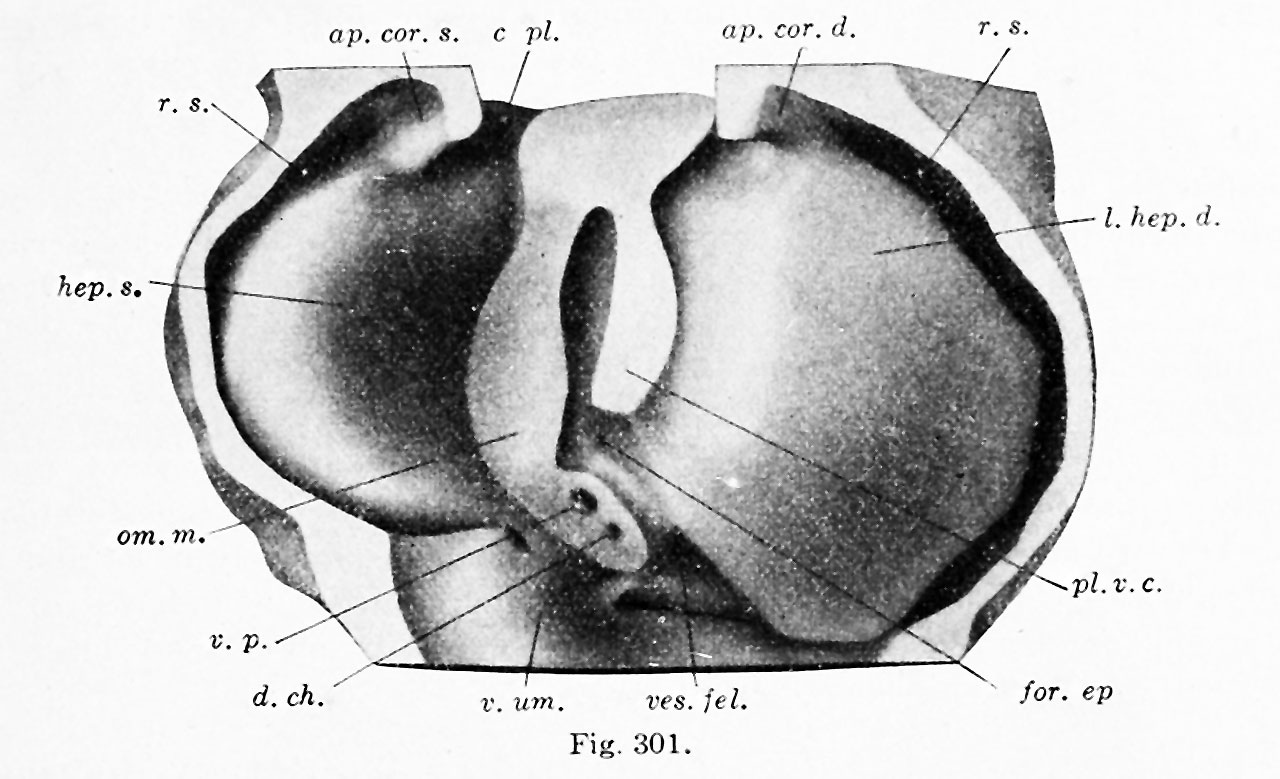 | 1,280 × 779 (140 KB) | Z8600021 (talk | contribs) |
You cannot overwrite this file.
File usage
The following 3 pages use this file: