File:Keibel Mall 2 169.jpg

Original file (634 × 800 pixels, file size: 97 KB, MIME type: image/jpeg)
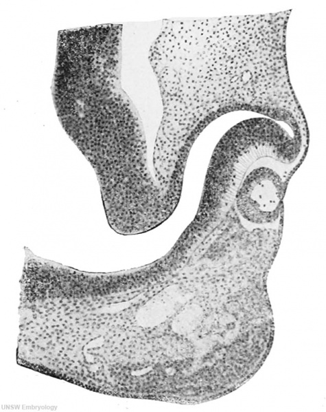
Fig. 169. Section through the optic anlage of an embryo of 7 mm
From a drawing kindly furnished me by Professor Hochstetter of Vienna. X 100. For explanation see text.
| Embryology - 16 Apr 2024 |
|---|
| Google Translate - select your language from the list shown below (this will open a new external page) |
|
العربية | català | 中文 | 中國傳統的 | français | Deutsche | עִברִית | हिंदी | bahasa Indonesia | italiano | 日本語 | 한국어 | မြန်မာ | Pilipino | Polskie | português | ਪੰਜਾਬੀ ਦੇ | Română | русский | Español | Swahili | Svensk | ไทย | Türkçe | اردو | ייִדיש | Tiếng Việt These external translations are automated and may not be accurate. (More? About Translations) |
Felix W. The development of the urinogenital organs. In Keibel F. and Mall FP. Manual of Human Embryology II. (1912) J. B. Lippincott Company, Philadelphia. pp 752-979.
| Historic Disclaimer - information about historic embryology pages |
|---|
 |
Cite this page: Hill, M.A. (2024, April 16) Embryology Keibel Mall 2 169.jpg. Retrieved from https://embryology.med.unsw.edu.au/embryology/index.php/File:Keibel_Mall_2_169.jpg
- © Dr Mark Hill 2024, UNSW Embryology ISBN: 978 0 7334 2609 4 - UNSW CRICOS Provider Code No. 00098G
File history
Click on a date/time to view the file as it appeared at that time.
| Date/Time | Thumbnail | Dimensions | User | Comment | |
|---|---|---|---|---|---|
| current | 10:16, 21 February 2014 | | 634 × 800 (97 KB) | Z8600021 (talk | contribs) | ==Fig. 169. Section through the optic anlage of an embryo of 7 mm== From a drawing kindly furnished me by Professor Hochstetter of Vienna. X 100. For explanation see text. {{Human Embryology Manual 2 19}} |
You cannot overwrite this file.
File usage
The following page uses this file: