File:Keibel Mall 2 168.jpg
Original file (583 × 1,000 pixels, file size: 65 KB, MIME type: image/jpeg)
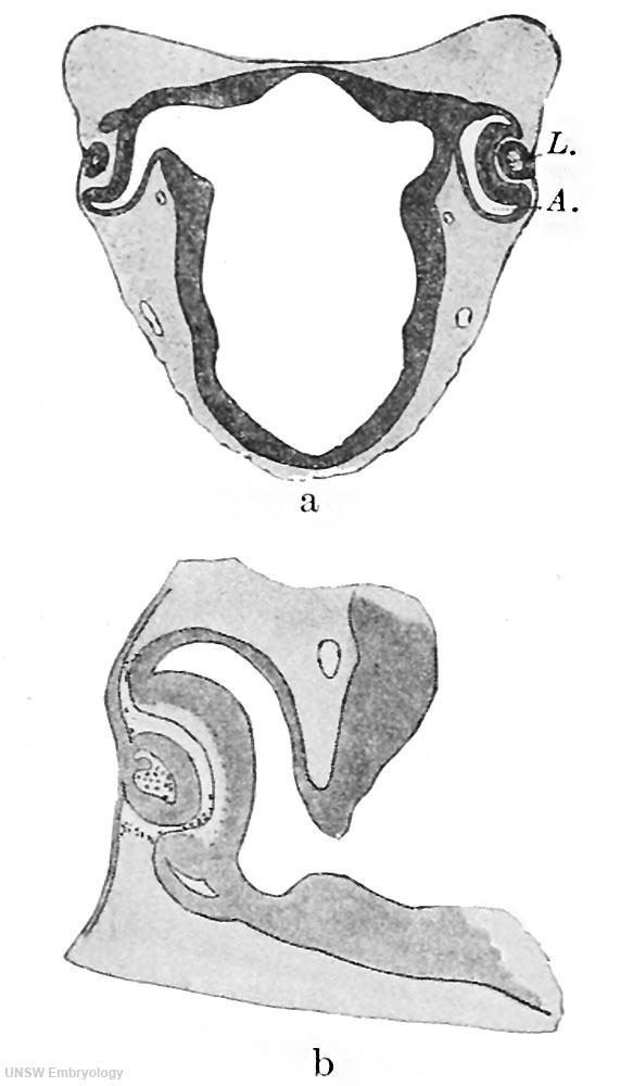
Fig. 168 a and b.Section through the anterior part of the head of a human embryo of 6.25 mm
a: Section through the anterior part of the head of a human embryo of 6.25 mm., passing through the optic cup and the lens vesicle which is being constricted off from the epidermis. In the interior of the lens vesicle there is a mass of degenerated cells, shown more distinctly in Fig. b. a X20; b X50.
(From the Normentafel of Keibel and Elze, Fig. I8 b and d.)
| Embryology - 25 Apr 2024 |
|---|
| Google Translate - select your language from the list shown below (this will open a new external page) |
|
العربية | català | 中文 | 中國傳統的 | français | Deutsche | עִברִית | हिंदी | bahasa Indonesia | italiano | 日本語 | 한국어 | မြန်မာ | Pilipino | Polskie | português | ਪੰਜਾਬੀ ਦੇ | Română | русский | Español | Swahili | Svensk | ไทย | Türkçe | اردو | ייִדיש | Tiếng Việt These external translations are automated and may not be accurate. (More? About Translations) |
Felix W. The development of the urinogenital organs. In Keibel F. and Mall FP. Manual of Human Embryology II. (1912) J. B. Lippincott Company, Philadelphia. pp 752-979.
| Historic Disclaimer - information about historic embryology pages |
|---|
 |
Cite this page: Hill, M.A. (2024, April 25) Embryology Keibel Mall 2 168.jpg. Retrieved from https://embryology.med.unsw.edu.au/embryology/index.php/File:Keibel_Mall_2_168.jpg
- © Dr Mark Hill 2024, UNSW Embryology ISBN: 978 0 7334 2609 4 - UNSW CRICOS Provider Code No. 00098G
File history
Click on a date/time to view the file as it appeared at that time.
| Date/Time | Thumbnail | Dimensions | User | Comment | |
|---|---|---|---|---|---|
| current | 10:17, 21 February 2014 | | 583 × 1,000 (65 KB) | Z8600021 (talk | contribs) | ==Fig. 168 a and b.Section through the anterior part of the head of a human embryo of 6.25 mm== a: Section through the anterior part of the head of a human embryo of 6.25 mm., passing through the optic cup and the lens vesicle which is being constrict... |
You cannot overwrite this file.
File usage
The following page uses this file: