File:Keibel Mall 291.jpg
Keibel_Mall_291.jpg (800 × 421 pixels, file size: 54 KB, MIME type: image/jpeg)
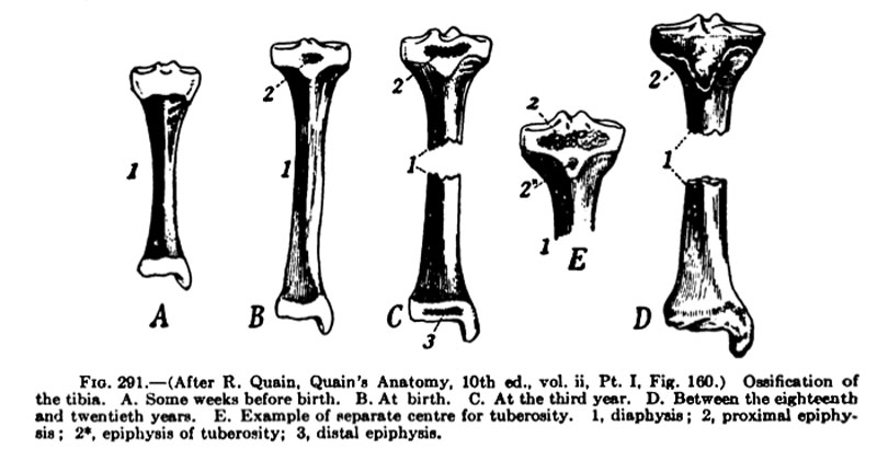
Fig. 291 Ossification of the Tibia
After R. Quain, Quain's Anatomy, 10th ed., vol. ii, Pt. I. Fig. 160.
- A - Some weeks before birth.
- B - At birth.
- C - At the third year.
- D - Betweoi the eighteenth and twentieth years.
- E - Example of separate centre for tuberosity.
1, diaphysis ; 2, proximal epiphysis; 2*, epiphysis of tuberosity; 3, distal epiphysis.
The earliest appearance of the tarsal cartilages is found in an embryo about 14 mm. long (Fig. 276). Toward the end of the second month these cartilages become much more distinct (Fig. 277). By the middle of the third month the cartilages of the foot have a form distinctly corresponding to the adult. The similarity is still better marked at the end of the third month (Fig. 278).
- Limb Images: 274-278 Spinal Column and Lower Limb | 279-284 Lower Limb | 285-288 Knee | 289 Os Coxae | 290 Femur | 291 Tibia | 292 Fibula | 293 Foot | 294 | 295 | 296 | 297 | 298-299 | 300 Forearm and Hand | 301 Upper Limb Joints | 302 Clavicle | Upper Limb Ossification 1 | Upper Limb Ossification 2 | Bone Development Timeline
- Skeleton and Connective Tissues: Connective Tissue Histogenesis | Skeletal Morphogenesis | Chorda Dorsalis | Vertebral Column and Thorax | Limb Skeleton | Skull Hyoid Bone Larynx
- KM Figure Links: The Germ Cells | Segmentation | First Primitive Segment | Gastrulation | External Form | Placenta | Axial Skeleton | Limb Skeleton | Skull | Muscular System
| Historic Disclaimer - information about historic embryology pages |
|---|
 |
Glossary Links
- Glossary: A | B | C | D | E | F | G | H | I | J | K | L | M | N | O | P | Q | R | S | T | U | V | W | X | Y | Z | Numbers | Symbols | Term Link
Cite this page: Hill, M.A. (2024, April 25) Embryology Keibel Mall 291.jpg. Retrieved from https://embryology.med.unsw.edu.au/embryology/index.php/File:Keibel_Mall_291.jpg
- © Dr Mark Hill 2024, UNSW Embryology ISBN: 978 0 7334 2609 4 - UNSW CRICOS Provider Code No. 00098G
File history
Click on a date/time to view the file as it appeared at that time.
| Date/Time | Thumbnail | Dimensions | User | Comment | |
|---|---|---|---|---|---|
| current | 10:35, 27 August 2012 | | 800 × 421 (54 KB) | Z8600021 (talk | contribs) | ==Fig. 291 Human Embryo Skeleton== {{KM Skeleton}} {{Keibel_Mall Images}} |
You cannot overwrite this file.