File:Jenkins010-012.jpg
From Embryology
Size of this preview: 800 × 597 pixels. Other resolution: 1,200 × 896 pixels.
Original file (1,200 × 896 pixels, file size: 193 KB, MIME type: image/jpeg)
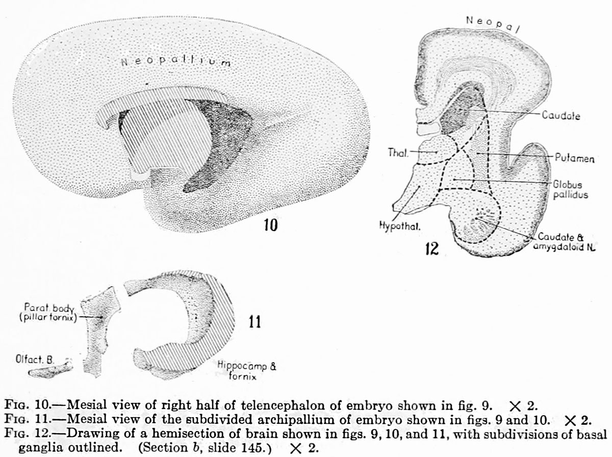
Fig. 10. Mesial view of right half of telencephalon of embryo shown in fig. 9
Fig. 11. Mesial view of the subdivided archipallium of embryo shown in figs. 9 and 10
Fig. 12. Drawing of a hemisection of brain shown in figs. 9, 10, and 11, with subdivisions of basal ganglia outlined. (Section 6, slide 145.)
all originally X 2.
File history
Click on a date/time to view the file as it appeared at that time.
| Date/Time | Thumbnail | Dimensions | User | Comment | |
|---|---|---|---|---|---|
| current | 08:08, 15 April 2015 | | 1,200 × 896 (193 KB) | Z8600021 (talk | contribs) | |
| 18:09, 15 February 2011 |  | 729 × 506 (51 KB) | S8600021 (talk | contribs) | ==Fig. 10. Mesial view of right half of telencephalon of embryo shown in fig. 9== ==Fig. 11. Mesial view of the subdivided archipallium of embryo shown in figs. 9 and 10== ==Fig. 12. Drawing of a hemisection of brain shown in figs. 9, 10, and 11, with |
You cannot overwrite this file.
File usage
The following page uses this file: