File:Integumentary- sebaceous gland histology 02.jpg
Integumentary-_sebaceous_gland_histology_02.jpg (400 × 500 pixels, file size: 150 KB, MIME type: image/jpeg)
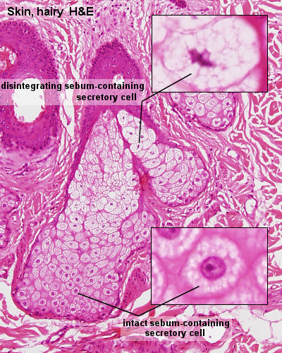
Sebaceous Gland Histology
(Stain - Haematoxylin Eosin)
Sebaceous glands empty their secretory product into the upper parts of the hair follicles, and together they form a pilosebaceous unit.
Sebaceous glands are also found in some of the areas where no hair is present; lips, oral surfaces of the cheeks and external genitalia.
Sebaceous glands are as a rule simple and branched. The secretory portion consists of alveoli. Basal cells in the outermost layer of the alveolus are flattened. Basal cells are mitotically active. Some of the new cells will replenish the pool of basal cells, while the remaining cells are displaced towards the centre of the alveolus as more cells are generated by the basal cells. The secretory cells will gradullay accumulate lipids and grow in size. Finally their nuclei disintegrate, and the cells rupture. The resulting secretory product of lipids and the constituents of the disintegrating cell is a holocrine secretion.
The lipid secretion of the sebaceous glands in humans is unclear. The secretion has no softening effect on the skin and only very limited antibacterial and antifungoid activity.
Clinically the sebaceous glands are important in that they are liable to infections, particularly during puberty. Template:Gland Histology
- Hair Links: Follicle in skin 1 | Follicle in skin 2 | Follicle label 1 | Follicle label 2 | Sebaceous gland 1 | Sebaceous gland 2
Links: Histology | Histology Stains | Blue Histology images copyright Lutz Slomianka 1998-2009. The literary and artistic works on the original Blue Histology website may be reproduced, adapted, published and distributed for non-commercial purposes. See also the page Histology Stains.
Cite this page: Hill, M.A. (2024, April 19) Embryology Integumentary- sebaceous gland histology 02.jpg. Retrieved from https://embryology.med.unsw.edu.au/embryology/index.php/File:Integumentary-_sebaceous_gland_histology_02.jpg
- © Dr Mark Hill 2024, UNSW Embryology ISBN: 978 0 7334 2609 4 - UNSW CRICOS Provider Code No. 00098G
File history
Click on a date/time to view the file as it appeared at that time.
| Date/Time | Thumbnail | Dimensions | User | Comment | |
|---|---|---|---|---|---|
| current | 12:20, 13 October 2010 | | 400 × 500 (150 KB) | S8600021 (talk | contribs) | ==Sebaceous Gland Histology== Sebaceous glands empty their secretory product into the upper parts of the hair follicles, and together they form a pilosebaceous unit. Sebaceous glands are also found in some of the areas where no hair is present; lips, or |
You cannot overwrite this file.