File:Images of congenital hereditary cataracts due to mutations of crystallin genes.png
Images_of_congenital_hereditary_cataracts_due_to_mutations_of_crystallin_genes.png (594 × 190 pixels, file size: 118 KB, MIME type: image/png)
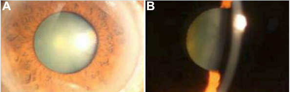
Slit-lamp photographs of the eye of the proband. Slit lamp photographs of the eye of the proband (III:3). A: Front view of the eye of the proband, showing cataract phenotype. B: Slit lamp view of the len of the proband. Lens opacities were mainly located in the nuclear area of lenses as well as in the embryonal and fetal areas.
Copyright notice This is an open-access article distributed under the terms of the Creative Commons Attribution License, which permits unrestricted use, distribution, and reproduction in any medium, provided the original work is properly cited.
File history
Click on a date/time to view the file as it appeared at that time.
| Date/Time | Thumbnail | Dimensions | User | Comment | |
|---|---|---|---|---|---|
| current | 17:48, 16 September 2012 | 594 × 190 (118 KB) | Z3331330 (talk | contribs) | Slit-lamp photographs of the eye of the proband. Slit lamp photographs of the eye of the proband (III:3). A: Front view of the eye of the proband, showing cataract phenotype. B: Slit lamp view of the len of the proband. Lens opacities were mainly located |
You cannot overwrite this file.
File usage
The following 3 pages use this file: