File:Hydrocephalus.jpg
Hydrocephalus.jpg (320 × 432 pixels, file size: 29 KB, MIME type: image/jpeg)
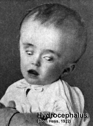
Hydrocephalus
(historic image from Hess, 1922)
This can be a defect of cerebrospinal fliud (CSF) flow, leading to enlarged ventricles and head, separated skull cranial sutures and fontanelles. Obstruction of CSF flow can occur at any time (prenatally or postnatally) and leads to accumulation of within the ventricles. The time of onset will have different effects and should be compared to the equilivant neurological events that are occuring.
Ventricular obstruction usually occurs at the level of the cerebral aqueduct (narrowest site), but can occur elsewhere, and can also be caused by a viral infection.
Links: Congenital Hydrocephalus
Cite this page: Hill, M.A. (2024, April 18) Embryology Hydrocephalus.jpg. Retrieved from https://embryology.med.unsw.edu.au/embryology/index.php/File:Hydrocephalus.jpg
- © Dr Mark Hill 2024, UNSW Embryology ISBN: 978 0 7334 2609 4 - UNSW CRICOS Provider Code No. 00098G
File history
Click on a date/time to view the file as it appeared at that time.
| Date/Time | Thumbnail | Dimensions | User | Comment | |
|---|---|---|---|---|---|
| current | 11:56, 26 January 2010 | | 320 × 432 (29 KB) | S8600021 (talk | contribs) | Hydrocephalus (historic image from Hess, 1922) This is a defect of cerebrospinal fliud (CSF) flow, leading to enlarged ventricles and head, separated skull cranial sutures and fontanelles. Obstruction of CSF flow can occur at any time (prenatally or post |
You cannot overwrite this file.
File usage
The following 15 pages use this file:
- 2010 BGD Practical 12 - Abnormalities
- 2010 BGD Tutorial - Applied Embryology and Teratology
- 2010 Lecture 1
- 2011 Lab 12 - Abnormalities
- ANAT2341 Lab 12 - Abnormalities
- Abnormal Development - Australian Statistics
- Lecture - 2011 Course Introduction
- Lecture - 2012 Course Introduction
- Lecture - 2013 Course Introduction
- Lecture - 2014 Course Introduction
- Lecture - 2015 Course Introduction
- Maternal-Fetal Medicine Trainees - Renal
- Talk:2010 BGD Tutorial - Applied Embryology and Teratology
- Talk:Lecture - 2016 Course Introduction
- Talk:Lecture - Renal Development