File:Huntington Disease patient and control MRI.gif: Difference between revisions
(Figure samples of coronal and sagittal MR images showing outlines for caudate, putamen, cerebral and cerebellar volumes in a patient with Huntington’s disease (top) and a normal control (bottom). http://www.scielo.br/scielo.php?script=sci_arttext&pid=S) |
No edit summary |
||
| Line 3: | Line 3: | ||
http://www.scielo.br/scielo.php?script=sci_arttext&pid=S0004-282X2006000100002&lng=en&nrm=iso&tlng=en | http://www.scielo.br/scielo.php?script=sci_arttext&pid=S0004-282X2006000100002&lng=en&nrm=iso&tlng=en | ||
==References== | |||
<pubmed>16622544</pubmed> | |||
< | |||
Attribution-NonCommercial 3.0 Unported (CC BY-NC 3.0) | Attribution-NonCommercial 3.0 Unported (CC BY-NC 3.0) | ||
You are free: | You are free: | ||
* to Share — to copy, distribute and transmit the work | |||
* to Remix — to adapt the work | |||
Under the following conditions: | Under the following conditions: | ||
* Attribution — You must attribute the work in the manner specified by the author or licensor (but not in any way * that suggests that they endorse you or your use of the work). | |||
Noncommercial — You may not use this work for commercial purposes. | |||
Notice — For any reuse or distribution, you must make clear to others the license terms of this work. The best way to do this is with a link to this web page. | |||
{{Template:2011 Student Image}} | {{Template:2011 Student Image}} | ||
Revision as of 10:07, 11 September 2011
Figure samples of coronal and sagittal MR images showing outlines for caudate, putamen, cerebral and cerebellar volumes in a patient with Huntington’s disease (top) and a normal control (bottom).
References
<pubmed>16622544</pubmed>
Attribution-NonCommercial 3.0 Unported (CC BY-NC 3.0)
You are free:
- to Share — to copy, distribute and transmit the work
- to Remix — to adapt the work
Under the following conditions:
- Attribution — You must attribute the work in the manner specified by the author or licensor (but not in any way * that suggests that they endorse you or your use of the work).
Noncommercial — You may not use this work for commercial purposes.
Notice — For any reuse or distribution, you must make clear to others the license terms of this work. The best way to do this is with a link to this web page.
- Note - This image was originally uploaded as part of a student project and may contain inaccuracies in either description or acknowledgements. Students have been advised in writing concerning the reuse of content and may accidentally have misunderstood the original terms of use. If image reuse on this non-commercial educational site infringes your existing copyright, please contact the site editor for immediate removal.
Cite this page: Hill, M.A. (2024, April 23) Embryology Huntington Disease patient and control MRI.gif. Retrieved from https://embryology.med.unsw.edu.au/embryology/index.php/File:Huntington_Disease_patient_and_control_MRI.gif
- © Dr Mark Hill 2024, UNSW Embryology ISBN: 978 0 7334 2609 4 - UNSW CRICOS Provider Code No. 00098G
File history
Click on a date/time to view the file as it appeared at that time.
| Date/Time | Thumbnail | Dimensions | User | Comment | |
|---|---|---|---|---|---|
| current | 17:58, 10 September 2011 | 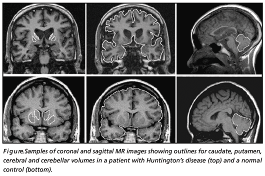 | 530 × 352 (139 KB) | Z3290379 (talk | contribs) | Figure samples of coronal and sagittal MR images showing outlines for caudate, putamen, cerebral and cerebellar volumes in a patient with Huntington’s disease (top) and a normal control (bottom). http://www.scielo.br/scielo.php?script=sci_arttext&pid=S |
You cannot overwrite this file.
File usage
The following 2 pages use this file: