File:Human zygote two pronuclei 03.jpg
Human_zygote_two_pronuclei_03.jpg (503 × 477 pixels, file size: 33 KB, MIME type: image/jpeg)
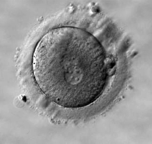
Human Early Zygote
- Early zygote observed after 18 to 20 hours after insemination.
- Zygote showing the two unfused pronuclei (male and female) containing nucleolar precursor bodies.
- Polar bodies are seen (3 o'clock position) at the periphery separating the zygote plasma membrane from the surrounding zone pellucida.
- Zygote Image Links: Image - Early zygote | Image - Early zygote labelled | Image - zygote 1 | Image - zygote 2 | Image - zygote 3 | Image - zygote 2 labeled | Fertilization | Zygote | Carnegie stage 1 | Category:Zygote
Reference
Copyright
© 2010 Nicoli et al; licensee BioMed Central Ltd. This is an Open Access article distributed under the terms of the Creative Commons Attribution License (http://creativecommons.org/licenses/by/2.0), which permits unrestricted use, distribution, and reproduction in any medium, provided the original work is properly cited.
Original File name: Figure 2. 1477-7827-8-77-2-l.jpg
Original magnification ×400, for the paper publication not the online image.
The original figure (B) was extracted from the full panel image and modified using Photoshop with the addition of labels:
- (B) 2PN centralized and juxtaposed, NPBs dispersed in the 2PN, perpendicular PB alignment.
(A) 2PN centralized and juxtaposed, NPBs aligned on the side of the 2PN, longitudinal PB alignment. (B) 2PN centralized and juxtaposed, NPBs dispersed in the 2PN, perpendicular PB alignment. (C) 2PN centralized and juxtaposed, NPBs non polarized with dispersed or not completely aligned NPBs in the 2PN, longitudinal PB alignment. (D) 2PN centralized and juxtaposed, NPBs aligned on the side of the 2PN, neither longitudinal nor perpendicular PB alignment. Black narrows indicate the PBs. (A) and (B) were examples of zygotes included in Pattern 1 group, while (C) and (D) were examples of zygotes included in Pattern 2 group.
File history
Click on a date/time to view the file as it appeared at that time.
| Date/Time | Thumbnail | Dimensions | User | Comment | |
|---|---|---|---|---|---|
| current | 16:27, 19 July 2010 | | 503 × 477 (33 KB) | S8600021 (talk | contribs) | Zygote showing different distribution of nucleolar precursor bodies (NPB) in the two pronuclei (2PN) and different PB aligment (Original magnification ×400, for the paper publication not the online image). Zygote observed after 18-20 hours after insemina |
You cannot overwrite this file.
File usage
The following page uses this file: