File:Human week 10 fetus 26.jpg: Difference between revisions
mNo edit summary |
mNo edit summary |
||
| Line 6: | Line 6: | ||
Loops of the midgut can be seen lying outside the ventral body wall (midgut herniation) but still connected by their mesentery to the posterior body wall. Developing villi can be seen in cross-sections of the midgut. The extensive underlying submucosa is visible and the outer muscularis layer is developing. | Loops of the midgut can be seen lying outside the ventral body wall (midgut herniation) but still connected by their mesentery to the posterior body wall. Developing villi can be seen in cross-sections of the midgut. The extensive underlying submucosa is visible and the outer muscularis layer is developing. | ||
Mesentery is seen attached to some of the midgut loops, but in fact forms a continuous connection to the length of the entire midgut, just not visible in this section. Note the may vessels lying within the mesentery. | |||
Hindgut lying within the body peritoneal cavity has a different histological appearance from the mid-gut. | Hindgut lying within the body peritoneal cavity has a different histological appearance from the mid-gut. | ||
Revision as of 16:58, 25 May 2016
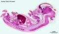
Human Female Fetus - Midgut Herniation (10 week)
Large image version of plane D, close to midline (Stain - Haematoxylin Eosin) 0.5 mm scale bar
Loops of the midgut can be seen lying outside the ventral body wall (midgut herniation) but still connected by their mesentery to the posterior body wall. Developing villi can be seen in cross-sections of the midgut. The extensive underlying submucosa is visible and the outer muscularis layer is developing.
Mesentery is seen attached to some of the midgut loops, but in fact forms a continuous connection to the length of the entire midgut, just not visible in this section. Note the may vessels lying within the mesentery.
Hindgut lying within the body peritoneal cavity has a different histological appearance from the mid-gut.
- Human Female Fetus (week 10)
Sagittal Section (plane D)

Pituitary and Lamina Terminalis

Olfactory Nerve

Atlas and Axis

Sacrum

Oral Cavity

Epiglottis

Heart

Spleen

Midgut Herniation

Midgut Herniation (label)

Pelvic Region

Pelvic Region (label)


Related Images
Fetus (week 10) Planes A (most lateral), B (lateral), C (medial) and D (midline) from lateral towards the midline.
- Human Fetus - most lateral | lateral | medial | midline
- Head - most lateral | lateral | medial | midline
- Cerebellum - most lateral | lateral | medial | midline
- Urogenital Unlabelled - most lateral | lateral | medial | midline
- Urogenital Labelled - most lateral | lateral | medial | midline
- Large Images - midline
- Image Source: UNSW Embryology, no reproduction without permission.
File history
Click on a date/time to view the file as it appeared at that time.
| Date/Time | Thumbnail | Dimensions | User | Comment | |
|---|---|---|---|---|---|
| current | 10:15, 1 May 2016 | 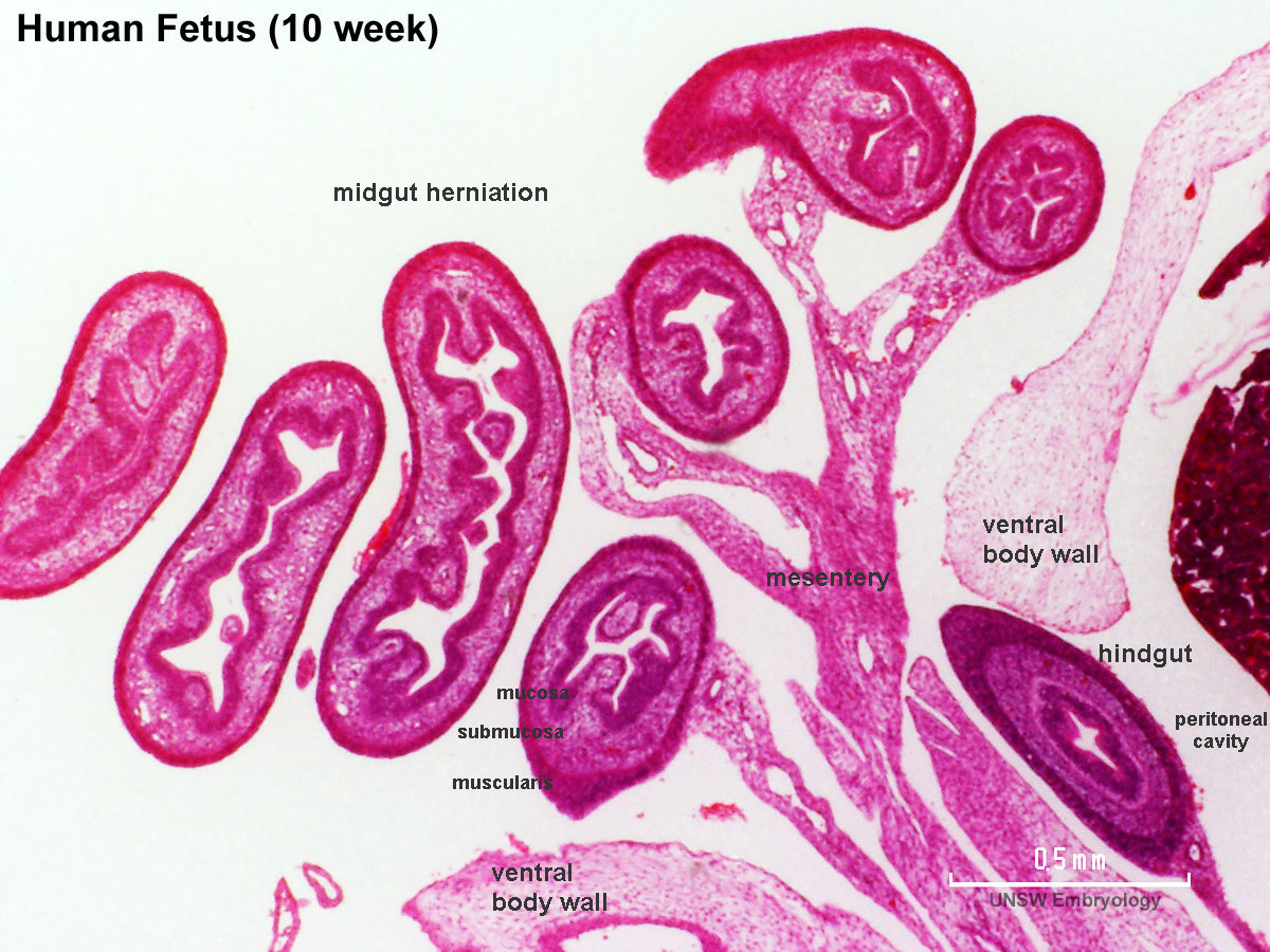 | 1,200 × 900 (262 KB) | Z8600021 (talk | contribs) |
You cannot overwrite this file.
File usage
The following 18 pages use this file:
- BGDA Practical 12 - Embryo to Fetus
- BGDB Gastrointestinal - Activity 2
- BGDB Gastrointestinal - Fetal
- Foundations Practical - Week 9 to 36
- File:Human week 10 fetus 01.jpg
- File:Human week 10 fetus 03.jpg
- File:Human week 10 fetus 04.jpg
- File:Human week 10 fetus 05.jpg
- File:Human week 10 fetus 06.jpg
- File:Human week 10 fetus 07.jpg
- File:Human week 10 fetus 08.jpg
- File:Human week 10 fetus 09.jpg
- File:Human week 10 fetus 10.jpg
- File:Human week 10 fetus 11.jpg
- File:Human week 10 fetus 12.jpg
- File:Human week 10 fetus 23.jpg
- File:Human week 10 fetus 26.jpg
- Template:Human Female Fetus Week 10 gallery