File:Human week 10 fetus 23.jpg: Difference between revisions
mNo edit summary |
mNo edit summary |
||
| Line 11: | Line 11: | ||
[[Renal System Development|'''Renal Development''']] | [[Renal System Development|'''Renal Development''']] | ||
The urinary bladder | The urinary bladder, from the urogenital sinus that in turn developed from the cloaca, is surrounded by the developing detrusor muscle. A ureter and the allantois can be seen extending from the top of the bladder. | ||
[[Gastrointestinal Tract Development|'''Gastrointestinal Development''']] | |||
Behind the developing uterus the rectum formed from hindgut is cut in longitudinal section. | |||
[[Musculoskeletal System Development|'''Musculoskeletal Development''']] | |||
On the ventral body wall the inguinal cone lies beneath the midgut herniation and beneath this is the pubic symphysis of the developing pelvis. Behind the rectum the vertebral column and intervertebral disc reigns can be seen. Note the vertebral bodies are cartilage and will ossify during the fetal period. | |||
{{Human Female Fetus Week 10 gallery}} | {{Human Female Fetus Week 10 gallery}} | ||
Latest revision as of 17:21, 25 May 2016
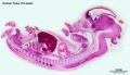
Human Female Fetus - Pelvic Region (10 week)
Large labelled image version of plane D, close to midline (Stain - Haematoxylin Eosin) 0.7 mm scale bar.
The genital tubercle can be seen at the top of the image and will contribute to the female external genitalia.
The early uterus forming from the paramesonephric duct lies in the peritoneal body cavity. Anterior to the uterus is the small uterovesical pouch (Dunn's pouch or pouch of Meiring), while posterior to the uterus is the larger rectouterine pouch (pouch of Douglas).
The urinary bladder, from the urogenital sinus that in turn developed from the cloaca, is surrounded by the developing detrusor muscle. A ureter and the allantois can be seen extending from the top of the bladder.
Behind the developing uterus the rectum formed from hindgut is cut in longitudinal section.
On the ventral body wall the inguinal cone lies beneath the midgut herniation and beneath this is the pubic symphysis of the developing pelvis. Behind the rectum the vertebral column and intervertebral disc reigns can be seen. Note the vertebral bodies are cartilage and will ossify during the fetal period.
- Human Female Fetus (week 10)
Sagittal Section (plane D)

Pituitary and Lamina Terminalis

Olfactory Nerve

Atlas and Axis

Sacrum

Oral Cavity

Epiglottis

Heart

Spleen

Midgut Herniation

Midgut Herniation (label)

Pelvic Region

Pelvic Region (label)


Related Images
Fetus (week 10) Planes A (most lateral), B (lateral), C (medial) and D (midline) from lateral towards the midline.
- Human Fetus - most lateral | lateral | medial | midline
- Head - most lateral | lateral | medial | midline
- Cerebellum - most lateral | lateral | medial | midline
- Urogenital Unlabelled - most lateral | lateral | medial | midline
- Urogenital Labelled - most lateral | lateral | medial | midline
- Large Images - midline
- Image Source: UNSW Embryology, no reproduction without permission.
File history
Click on a date/time to view the file as it appeared at that time.
| Date/Time | Thumbnail | Dimensions | User | Comment | |
|---|---|---|---|---|---|
| current | 21:01, 14 April 2016 | 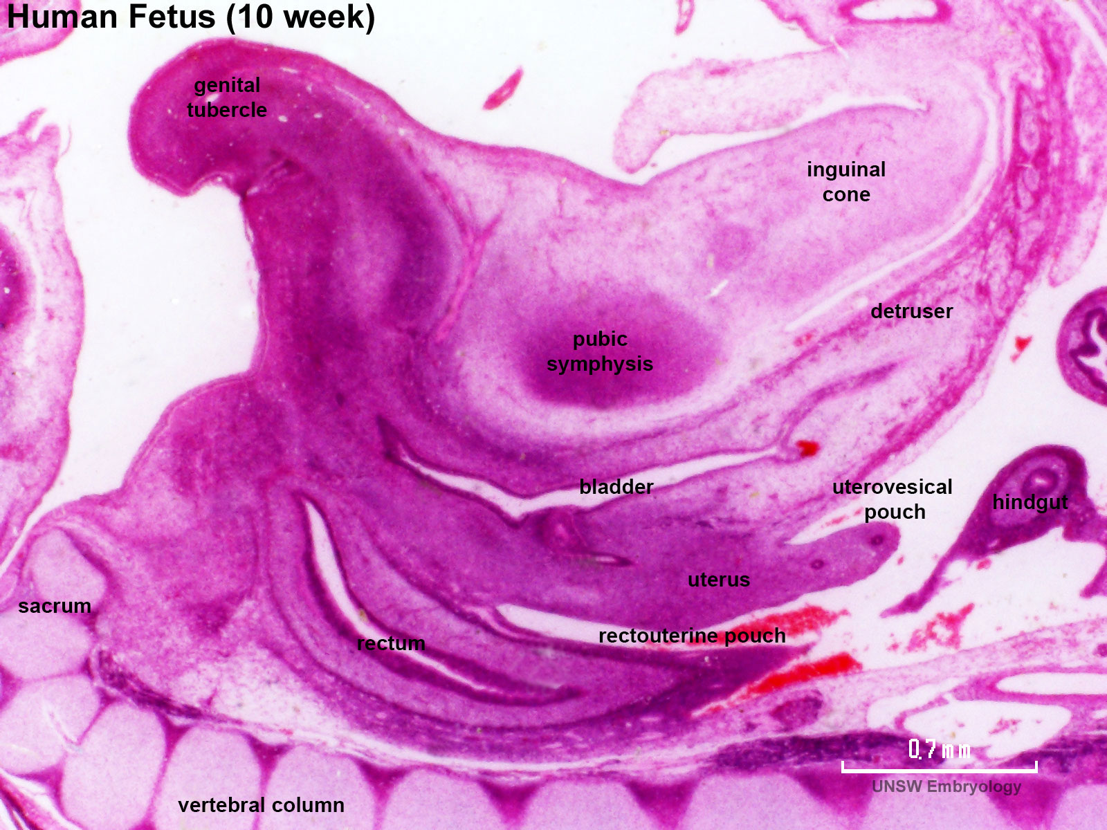 | 1,600 × 1,200 (393 KB) | Z8600021 (talk | contribs) |
You cannot overwrite this file.
File usage
The following 18 pages use this file:
- BGDA Practical 12 - Embryo to Fetus
- BGDB Gastrointestinal - Fetal
- Fetal Development - 10 Weeks
- Foundations Practical - Week 9 to 36
- File:Human week 10 fetus 01.jpg
- File:Human week 10 fetus 03.jpg
- File:Human week 10 fetus 04.jpg
- File:Human week 10 fetus 05.jpg
- File:Human week 10 fetus 06.jpg
- File:Human week 10 fetus 07.jpg
- File:Human week 10 fetus 08.jpg
- File:Human week 10 fetus 09.jpg
- File:Human week 10 fetus 10.jpg
- File:Human week 10 fetus 11.jpg
- File:Human week 10 fetus 12.jpg
- File:Human week 10 fetus 23.jpg
- File:Human week 10 fetus 26.jpg
- Template:Human Female Fetus Week 10 gallery