File:Human ovulation 06.jpg
Human_ovulation_06.jpg (734 × 583 pixels, file size: 82 KB, MIME type: image/jpeg)
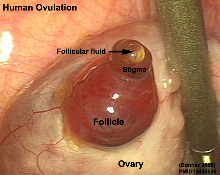
Human Ovulation
Laparoscopic observation of spontaneous human ovulation. A remarkably prominent vascular pattern was observed on the mature follicle (F, white arrows). A small follicular area called the stigma (S) was seen protruding like a reddish bleb from the follicular surface, with viscous yellow fluid (black arrows) evaginating outward into the peritoneal cavity. The viscous fluid probably carried with it the cumulus–oocyte complex, surrounded by several thousand small granulosa cells known as corona radiata.
Legend
- F - mature follicle, white arrows
- S - stigma
- black arrows - ovulating follicular fluid containing cumulus–oocyte surrounded by several thousand small granulosa cells
- Links: Full figure | Image 1 of 4 | Image 2 of 4 | Image 3 of 4 | Image 4 of 4 | Non-ovulating Ovary Image | Ovary Development | Uterus Development | Menstrual Cycle
Original file name: Figure 1 (cropped second image in set of four, removed original labels and relabeled)
File history
Click on a date/time to view the file as it appeared at that time.
| Date/Time | Thumbnail | Dimensions | User | Comment | |
|---|---|---|---|---|---|
| current | 11:54, 10 December 2010 | | 734 × 583 (82 KB) | S8600021 (talk | contribs) | ==Human Ovulation== Laparoscopic observation of spontaneous human ovulation. A remarkably prominent vascular pattern was observed on the mature follicle (F, white arrows). A small follicular area called the stigma (S) was seen protruding like a reddish b |
You cannot overwrite this file.