File:Human ovary postnatal growth.jpg: Difference between revisions
mNo edit summary |
|||
| Line 6: | Line 6: | ||
For comparison see also earlier Brazilian | For comparison see also earlier Brazilian{{#pmid:12034633|PMID12034633}} and Danish{{#pmid:8521066|PMID8521066}} data. | ||
:'''Links:''' | :'''Links:''' {{Ovary}} | {{Puberty}} | ||
==Reference== | ==Reference== | ||
{{#pmid:16891683}} | |||
<references/> | <references/> | ||
{{Footer}} | |||
[[Category:Human]] [[Category:Female]] [[Category:Ovary]] [[Category:Genital]] [[Category:Graph]] [[Category:Postnatal]] [[Category:Puberty]] | [[Category:Human]] [[Category:Female]] [[Category:Ovary]] [[Category:Genital]] [[Category:Graph]] [[Category:Postnatal]] [[Category:Puberty]][[Category:Timeline]] | ||
Latest revision as of 12:48, 25 March 2019
Human Ovary Postnatal Growth
Graph was constructed from Indian population data shown in "Table 1 – Ovarian Volume, Uterine Length and FCR According to Chronological Age (n = 214)"
- "The aim of our study was to determine the pattern of female reproductive organ growth in Indian girls from birth to 18 years of age and to correlate the uterine length, mean ovarian volume (MOV) and Fundo Cervical Ratio (FCR) with chronological age, bone age and pubertal breast staging. A cross sectional study was performed on 218 girls from birth to 18 years of age. Height, weight, stage of puberty, X-ray for bone age and transabdominal ultrasounds were performed on all girls. Higher chronological age, bone age and increase in breast stage significantly predicted higher MOV (P < 0.001) and higher uterine length (P < 0.001). The MOV, uterine length and FCR are positively correlated with chronological age, bone age, height, weight and breast staging. Data from present study may be useful in screening cases of precocious puberty and other disorders that may need further evaluation."
For comparison see also earlier Brazilian[1] and Danish[2] data.
Reference
Khadilkar VV, Khadilkar AV, Kinare AS, Tapasvi HS, Deshpande SS & Maskati GB. (2006). Ovarian and uterine ultrasonography in healthy girls between birth to 18 years. Indian Pediatr , 43, 625-30. PMID: 16891683
- ↑ Herter LD, Golendziner E, Flores JA, Becker E & Spritzer PM. (2002). Ovarian and uterine sonography in healthy girls between 1 and 13 years old: correlation of findings with age and pubertal status. AJR Am J Roentgenol , 178, 1531-6. PMID: 12034633 DOI.
- ↑ Holm K, Laursen EM, Brocks V & Müller J. (1995). Pubertal maturation of the internal genitalia: an ultrasound evaluation of 166 healthy girls. Ultrasound Obstet Gynecol , 6, 175-81. PMID: 8521066 DOI.
Cite this page: Hill, M.A. (2024, April 18) Embryology Human ovary postnatal growth.jpg. Retrieved from https://embryology.med.unsw.edu.au/embryology/index.php/File:Human_ovary_postnatal_growth.jpg
- © Dr Mark Hill 2024, UNSW Embryology ISBN: 978 0 7334 2609 4 - UNSW CRICOS Provider Code No. 00098G
File history
Click on a date/time to view the file as it appeared at that time.
| Date/Time | Thumbnail | Dimensions | User | Comment | |
|---|---|---|---|---|---|
| current | 16:41, 4 January 2011 | 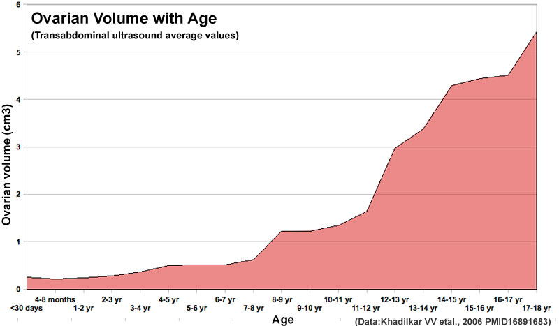 | 800 × 467 (40 KB) | S8600021 (talk | contribs) | ==Human Ovary Postnatal Growth== Graph was constructed from Indian population data shown in "TABLE I – Ovarian Volume, Uterine Length and FCR According to Chronological Age (n = 214)" ==Reference== <pubmed>16891683</pubmed> Category:Human [[Cat |
You cannot overwrite this file.
File usage
The following 3 pages use this file: