File:Human ovary - corpus luteum 21.jpg: Difference between revisions
(Z8600021 uploaded a new version of File:Human ovary - corpus luteum 21.jpg) |
(Z8600021 uploaded a new version of File:Human ovary - corpus luteum 21.jpg) |
(No difference)
| |
Latest revision as of 10:27, 6 May 2018
Human ovary - Corpus Luteum
Section through the middle of a human ovary at mid-luteal phase of the menstrual cycle after ovulation. (unlabeled image).
- Corpus luteum (white ring) shown at top left of ovary. Note the size and colour of the the corpus luteum ("yellow body") compared to the ovary and other internal structures. If implantation and pregnancy occurs, then the corpus luteum would also increase in size and hormonal function during the early pregnancy.
- white asterisks - possible older corpora lutea remnants.
- Corpus albicans (white arrow) shown at the bottom right of the ovary. Note the size and colour of the corpus albicans ("white body") compared to the corpus luteum and ovary. The corpus albicans is the degenerating non-functional form of the corpus luteum that "develops" from it if implantation and pregnancy do not occur.
- black asterisks - possible older corpora albicantia remnants.
Black arrows show the approximate cortex and medullary regions of the ovary.
- Links: labeled large image | large image | labeled small image | small image | Virtual Image | histology overview | Menstrual Cycle | Ovary Development | Corpus Luteum | Corpus Albicans | Category:Corpus Luteum
Image: Dr Ed Uthman (Houston, Texas) - other pathology images Original image has been resized and labeled.
Creative Commons Attribution-Share Alike 3.0 Unported
Cite this page: Hill, M.A. (2024, April 19) Embryology Human ovary - corpus luteum 21.jpg. Retrieved from https://embryology.med.unsw.edu.au/embryology/index.php/File:Human_ovary_-_corpus_luteum_21.jpg
- © Dr Mark Hill 2024, UNSW Embryology ISBN: 978 0 7334 2609 4 - UNSW CRICOS Provider Code No. 00098G
File history
Click on a date/time to view the file as it appeared at that time.
| Date/Time | Thumbnail | Dimensions | User | Comment | |
|---|---|---|---|---|---|
| current | 10:27, 6 May 2018 |  | 1,024 × 979 (89 KB) | Z8600021 (talk | contribs) | |
| 10:26, 6 May 2018 | 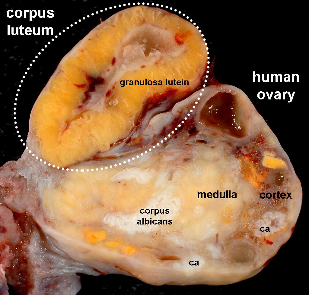 | 1,024 × 979 (89 KB) | Z8600021 (talk | contribs) | ||
| 10:42, 3 March 2014 | 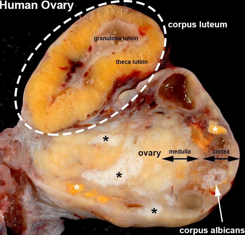 | 800 × 765 (133 KB) | Z8600021 (talk | contribs) | Added ovary labels. | |
| 10:15, 3 March 2014 | 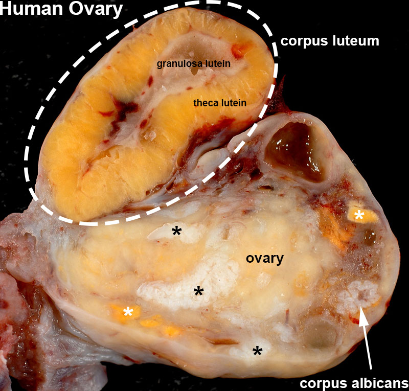 | 800 × 765 (131 KB) | Z8600021 (talk | contribs) | ==Human ovary - Corpus Luteum== {{Human ovary - corpus luteum links}} {{Ed Uthman}} Taken on July 17, 2008 Nikon D200 http://www.flickr.com/photos/euthman/2678061394/ [https://creativecommons.org/licenses/by-sa/3.0/ Creative Commons Attribution-S... |
You cannot overwrite this file.
File usage
The following file is a duplicate of this file (more details):
The following page uses this file: