File:Human heart SEM1.jpg: Difference between revisions
From Embryology
No edit summary |
(Z8600021 uploaded a new version of "File:Human heart SEM1.jpg") |
(No difference)
| |
Revision as of 13:50, 24 August 2014
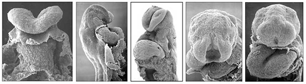
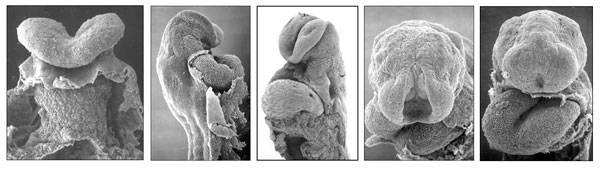
The Human Heart from day 21 to 25
Scanning electron micrograph images of the early human embryonic heart tube. Note the anterior body wall has been removed exposing the pericardial cavity in which the heart tube lies.
File history
Click on a date/time to view the file as it appeared at that time.
| Date/Time | Thumbnail | Dimensions | User | Comment | |
|---|---|---|---|---|---|
| current | 13:50, 24 August 2014 | 1,200 × 330 (47 KB) | Z8600021 (talk | contribs) | ||
| 21:21, 16 August 2009 | 600 × 177 (30 KB) | S8600021 (talk | contribs) | The Human Heart from day 10 to 25 |
You cannot overwrite this file.
File usage
The following 13 pages use this file:
- 2009 Lecture 7
- 2010 BGD Lecture - Development of the Embryo/Fetus 1
- 2010 Lecture 7
- ANAT2341 Lab 4 - Early Cardiovascular Development
- BGDA Lecture - Development of the Embryo/Fetus 1
- Cardiovascular System - Circulation Development
- Cardiovascular System - Coronary Circulation Development
- Cardiovascular System Development
- Fetal ECHO Meeting 2012
- Human Embryo SEM
- Lecture - Early Vascular Development
- RPAH Cardiac Embryology 2014
- User:Z3267024