File:Human embryonic renal branching 1.jpg: Difference between revisions
mNo edit summary |
mNo edit summary |
||
| Line 1: | Line 1: | ||
==Embryonic Human Renal Urothelial Branching Development== | ==Embryonic Human Renal Urothelial Branching Development== | ||
Renal Urothelial Branching Development at {{CS14}}, {{CS16}}, {{CS18}}, {{CS19}} and {{CS22}}. | Renal Urothelial Branching Development in embryos from the [[Kyoto Collection]] at carnegie stages: {{CS14}}, {{CS16}}, {{CS18}}, {{CS19}} and {{CS22}}. | ||
Development of the metanephroses and urinary collecting system during embryonic periods. Numbers indicate Carnegie stage. Scale bar = 0.5 mm. | Development of the metanephroses and urinary collecting system during embryonic periods. Numbers indicate Carnegie stage. Scale bar = 0.5 mm. | ||
{{Renal | |||
{{Renal Links}} | |||
===Reference=== | ===Reference=== | ||
| Line 14: | Line 15: | ||
Fig. 1. https://doi.org/10.1371/journal.pone.0203623.g001 Original figure resized, PMID added and converted to jpg format. | Fig. 1. https://doi.org/10.1371/journal.pone.0203623.g001 Original figure resized, PMID added and converted to jpg format. | ||
{{Footer}} | {{Footer}} | ||
[[Category:Renal]] | [[Category:Renal]][[Category:Carnegie Stage:14]][[Category:Carnegie Stage:16]][[Category:Carnegie Stage:18]][[Category:Carnegie Stage:19]][[Category:Carnegie Stage:22]][[Category:Kyoto Collection]] | ||
Revision as of 10:12, 18 January 2019
Embryonic Human Renal Urothelial Branching Development
Renal Urothelial Branching Development in embryos from the Kyoto Collection at carnegie stages: 14, 16, 18, 19 and 22.
Development of the metanephroses and urinary collecting system during embryonic periods. Numbers indicate Carnegie stage. Scale bar = 0.5 mm.
Reference
Ishiyama H, Ishikawa A, Kitazawa H, Fujii S, Matsubayashi J, Yamada S & Takakuwa T. (2018). Branching morphogenesis of the urinary collecting system in the human embryonic metanephros. PLoS ONE , 13, e0203623. PMID: 30192900 DOI.
Copyright
© 2018 Ishiyama et al. This is an open access article distributed under the terms of the Creative Commons Attribution License, which permits unrestricted use, distribution, and reproduction in any medium, provided the original author and source are credited.
Fig. 1. https://doi.org/10.1371/journal.pone.0203623.g001 Original figure resized, PMID added and converted to jpg format.
Cite this page: Hill, M.A. (2024, April 19) Embryology Human embryonic renal branching 1.jpg. Retrieved from https://embryology.med.unsw.edu.au/embryology/index.php/File:Human_embryonic_renal_branching_1.jpg
- © Dr Mark Hill 2024, UNSW Embryology ISBN: 978 0 7334 2609 4 - UNSW CRICOS Provider Code No. 00098G
File history
Click on a date/time to view the file as it appeared at that time.
| Date/Time | Thumbnail | Dimensions | User | Comment | |
|---|---|---|---|---|---|
| current | 10:07, 18 January 2019 | 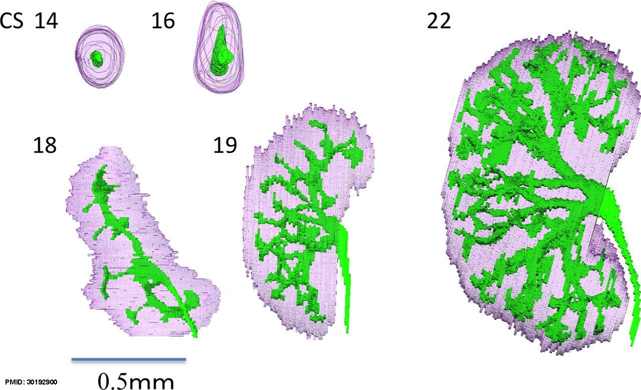 | 1,280 × 779 (236 KB) | Z8600021 (talk | contribs) | ==Embryonic Human Renal Urothelial Branching Development== Renal Urothelial Branching Development at {{CS14}} and {{CS22}}. Development of the metanephroses and urinary collecting system during embryonic periods. Numbers indicate Carnegie stage. Scal... |
You cannot overwrite this file.
File usage
The following page uses this file: