File:Human embryo thymus and parathyroid 01.jpg: Difference between revisions
mNo edit summary |
m (→Reference) |
||
| Line 20: | Line 20: | ||
===Reference=== | ===Reference=== | ||
{{#pmid:21203493}} | |||
Latest revision as of 23:10, 20 May 2019
Human Embryo Thymus and Parathyroid
Ectopic parathyroids are present from week 7 in the human embryo.
Whole-mount in situ hybridization for GCM2 (A–D, F, H–K) and whole-mounts embedded in paraffin and sectioned after in situ hybridization (E, G). Ages of embryos or dissected parathyroid/thymus primordia in the lower right corner of A, C, and F apply to the entire row; age in J also applies to K.
The entire thymus/parathyroid common primoridum is outlined in D, F, G, and H. In panels D–K, black arrows show presumptive primary parathyroids, small arrowheads indicate GCM2-positive clusters at the posterior end of the thymus, and red arrows show probable accessory parathyroids. White arrows in J and K show trailing GCM2-positive cells.
(A, B) Side (A) and frontal (B) views show GCM2 expression in the 3rd and 4th pharyngeal pouches in an early week 6 embryo (2/2 embryos).
(C–E) Images of whole-mount (C, D) and paraffin sectioned (E, section from D) early to mid week 7 dissected parathyroid/thymus common primordia showing GCM2 expressing cells present in the anterior (4/4 embryos) and posterior tip (1/4 embryos) of the common primordia. Small ectopic clusters away from the main cluster (*) are also often present by this stage.
(F) Image shows separation of parathyroids from the common primordia is occurring by late week 7 (right arrow; 1/1 embryo; the posterior parathyroid was present in only one of the two common primordia in this embryo).
(G) Paraffin section of whole mount shown in F. (H) Whole mount showing bilateral primordia (outlined) and carotid artery complex (red dashed line), with four main parathyroids and one small parathyroid cluster on the posterior tip of one thymic lobe.
(I, J) Thymic lobes and surrounding parathyroids from one side each of two separate early week 8 embryos show Gcm2 expression by three apparent primary parathyroids and a smaller accessory parathyroid (red arrow; 3/3 embryos; in each case the thymus/parathyroid primordium was examined from only one side of the embryo). The white arrow points to Gcm2 expressing cells that are still attached to the thymic domain.
(K) higher magnification of the region boxed in J. pt, parathyroid; th, thymus; p3, 3rd pharyngeal pouch; p4, 4th pharyngeal pouch.
Reference
Liu Z, Farley A, Chen L, Kirby BJ, Kovacs CS, Blackburn CC & Manley NR. (2010). Thymus-associated parathyroid hormone has two cellular origins with distinct endocrine and immunological functions. PLoS Genet. , 6, e1001251. PMID: 21203493 DOI.
Copyright
© 2010 Liu et al. This is an open-access article distributed under the terms of the Creative Commons Attribution License, which permits unrestricted use, distribution, and reproduction in any medium, provided the original author and source are credited.
Figure 4. Journal.pgen.1001251.g004.jpg
doi:info:doi/10.1371/journal.pgen.1001251.g004
File history
Click on a date/time to view the file as it appeared at that time.
| Date/Time | Thumbnail | Dimensions | User | Comment | |
|---|---|---|---|---|---|
| current | 17:32, 21 May 2012 | 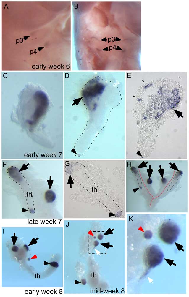 | 639 × 1,000 (58 KB) | Z8600021 (talk | contribs) | ==Human Embryo Thymus and Parathyroid== Ectopic parathyroids are present from week 7 in the human embryo Whole-mount in situ hybridization for GCM2 (A–D, F, H–K) and whole-mounts embedded in paraffin and sectioned after in situ hybridization (E, G). |
You cannot overwrite this file.
File usage
The following 2 pages use this file: