File:Human cochlea stria vascularis 01.jpg
Original file (1,854 × 1,806 pixels, file size: 754 KB, MIME type: image/jpeg)
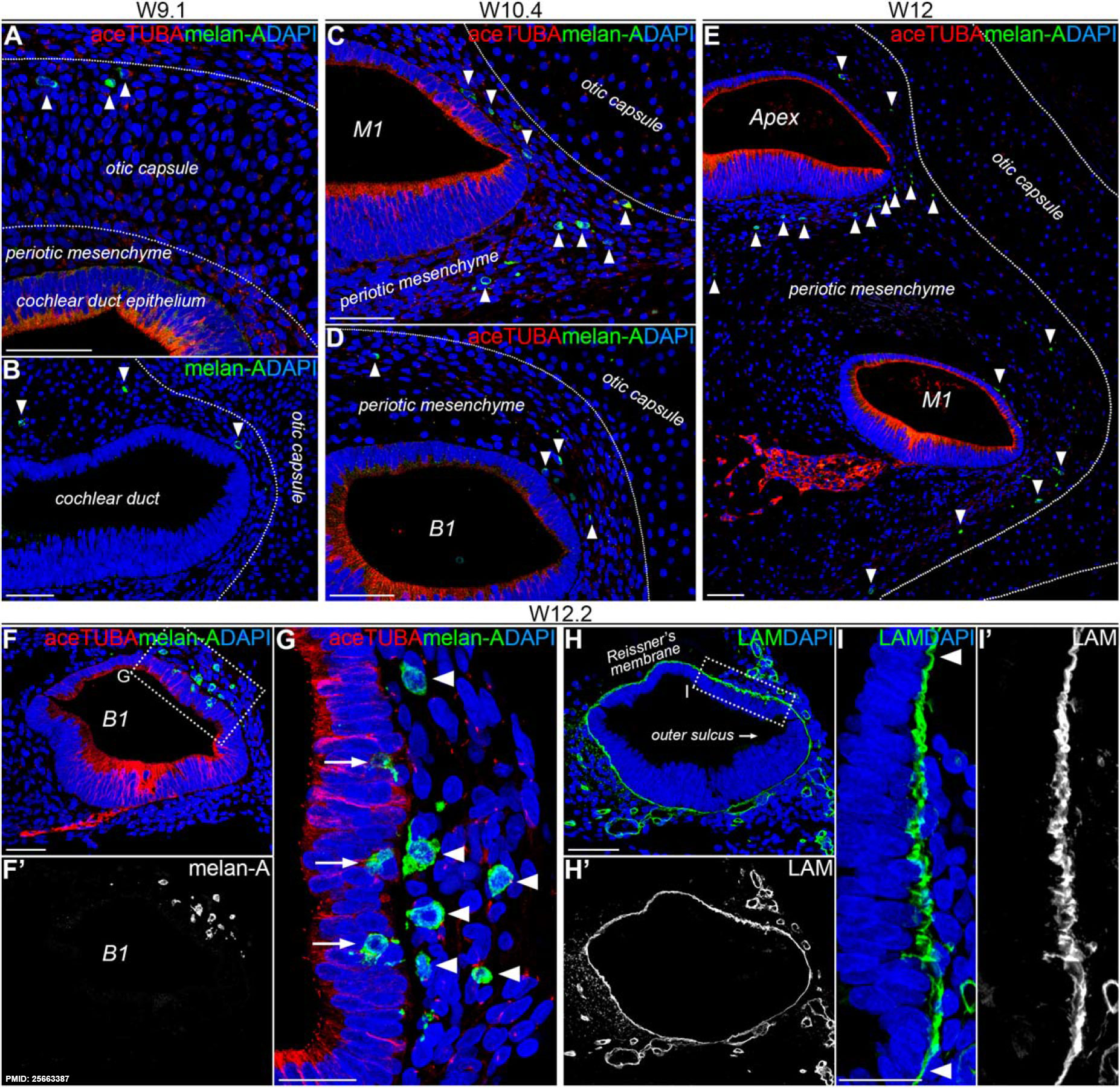
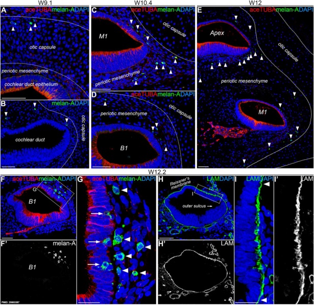
Migration of melanocytes into the stria vascularis of the human fetal cochlea
(A-B) A cochlea at W9.1 immunostained for acetylated tubulin (aceTUBA; red), melan-A (green) and DAPI (blue) (A) or melan-A (green) and DAPI (blue) (B). Arrowheads point to melan-A+ cochlear melanocytes. Scale bars = 50 µm. (C) The lower middle turn (M1) of a cochlea at W10.4 immunostained for aceTUBA (red), melan-A (green), and DAPI (blue). Scale bar = 50 µm. (D) The lower basal turn (B1) of a cochlea at W10.4 immunostained for aceTUBA (red), melan-A (green), and DAPI (blue). Scale bar = 50 µm. (E) The apical and lower middle turn of cochlea at W12 immunostained for aceTUBA (red), melan-A (green), and DAPI (blue). Scale bar = 50 µm. (F-F′) The lower basal turn of a cochlea at W12.2 immunostained for aceTUBA (red), melan-A (green), and DAPI (blue). The melan-A signal is shown separately in white in (F′). Scale bar = 50 µm. (G). Higher magnification of the outlined area in (F). Arrowheads point to melan-A+ melanocytes located in the periotic mesenchyme. Arrows point to melan-A+ melanocytes located in between the epithelial lining of the cochlear duct lateral wall (the future marginal cells of the stria vascularis). Scale bar = 20 µm. (H-H′). A similar section of the lower basal turn of a cochlea at W12.2 as depicted in (F), here immunostained for basement membrane protein laminin (LAM). The LAM signal is shown separately in white in (H′). Scale bar = 50 µm. (I-I′) Higher magnification of the developing stria vascularis outlined in (H). The arrowheads point to the smooth and continuous appearance of the basement membrane at the location of the bordering Reissner's membrane and the outer sulcus, as opposed to the irregular pattern observed in at the location of the future stria vascularis. The LAM signal is shown separately in white in (I′) Scale bar = 20 µm.
Reference
<pubmed>25663387</pubmed>| Dev Neurobiol.
Copyright
© 2015 The Authors Developmental Neurobiology Published by Wiley Periodicals, Inc. This is an open access article under the terms of the Creative Commons Attribution-NonCommercial License, which permits use, distribution and reproduction in any medium, provided the original work is properly cited and is not used for commercial purposes.
Figure 2. relabelled with pubmed ID
File history
Click on a date/time to view the file as it appeared at that time.
| Date/Time | Thumbnail | Dimensions | User | Comment | |
|---|---|---|---|---|---|
| current | 12:50, 11 April 2015 | | 1,854 × 1,806 (754 KB) | Z8600021 (talk | contribs) | ==Migration of melanocytes into the stria vascularis of the human fetal cochlea== (A-B) A cochlea at W9.1 immunostained for acetylated tubulin (aceTUBA; red), melan-A (green) and DAPI (blue) (A) or melan-A (green) and DAPI (blue) (B). Arrowheads point... |
You cannot overwrite this file.
File usage
The following 7 pages use this file: