File:Human cochlea stria vascularis 01.jpg: Difference between revisions
mNo edit summary |
mNo edit summary |
||
| (One intermediate revision by the same user not shown) | |||
| Line 11: | Line 11: | ||
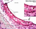
Scale bar = 20 µm. | Scale bar = 20 µm. | ||
{{Stria vascularis links}} | {{Stria vascularis links}} | ||
===Reference=== | ===Reference=== | ||
{{#pmid:25663387}} | |||
====Copyright==== | ====Copyright==== | ||
| Line 26: | Line 23: | ||
Figure 2. relabelled with pubmed ID | Figure 2. relabelled with pubmed ID | ||
{{Footer}} | |||
[[Category:Hearing]][[Category:Inner Ear]][[Category:Neural Crest]][[Category:Fluorescence]] | [[Category:Hearing]][[Category:Inner Ear]][[Category:Neural Crest]][[Category:Fluorescence]] | ||
Latest revision as of 10:39, 18 December 2018
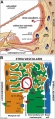
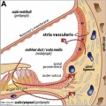
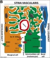
Migration of melanocytes into the stria vascularis of the human fetal cochlea
(A-B) A cochlea at W9.1 immunostained for acetylated tubulin (aceTUBA; red), melan-A (green) and DAPI (blue) (A) or melan-A (green) and DAPI (blue)
(B). Arrowheads point to melan-A+ cochlear melanocytes. Scale bars = 50 µm. (C) The lower middle turn (M1) of a cochlea at W10.4 immunostained for aceTUBA (red), melan-A (green), and DAPI (blue). Scale bar = 50 µm. (D) The lower basal turn (B1) of a cochlea at W10.4 immunostained for aceTUBA (red), melan-A (green), and DAPI (blue). Scale bar = 50 µm.
(E) The apical and lower middle turn of cochlea at W12 immunostained for aceTUBA (red), melan-A (green), and DAPI (blue). Scale bar = 50 µm. (F-F′) The lower basal turn of a cochlea at W12.2 immunostained for aceTUBA (red), melan-A (green), and DAPI (blue). The melan-A signal is shown separately in white in (F′). Scale bar = 50 µm.
(G). Higher magnification of the outlined area in (F). Arrowheads point to melan-A+ melanocytes located in the periotic mesenchyme. Arrows point to melan-A+ melanocytes located in between the epithelial lining of the cochlear duct lateral wall (the future marginal cells of the stria vascularis). Scale bar = 20 µm. (H-H′). A similar section of the lower basal turn of a cochlea at W12.2 as depicted in (F), here immunostained for basement membrane protein laminin (LAM). The LAM signal is shown separately in white in (H′). Scale bar = 50 µm. (I-I′) Higher magnification of the developing stria vascularis outlined in (H). The arrowheads point to the smooth and continuous appearance of the basement membrane at the location of the bordering Reissner's membrane and the outer sulcus, as opposed to the irregular pattern observed in at the location of the future stria vascularis. The LAM signal is shown separately in white in (I′)
Scale bar = 20 µm.
- Cochlea Links: stria vascular histology | stria vascularis 1 | stria vascularis 2 | stria vascularis 3 | human vascularis development | Neural Crest Development | Inner Ear Development
stria vascular histology
stria vascular 1
stria vascular 2
stria vascular 3
vascularis development


Reference
Locher H, de Groot JC, van Iperen L, Huisman MA, Frijns JH & Chuva de Sousa Lopes SM. (2015). Development of the stria vascularis and potassium regulation in the human fetal cochlea: Insights into hereditary sensorineural hearing loss. Dev Neurobiol , 75, 1219-40. PMID: 25663387 DOI.
Copyright
© 2015 The Authors Developmental Neurobiology Published by Wiley Periodicals, Inc. This is an open access article under the terms of the Creative Commons Attribution-NonCommercial License, which permits use, distribution and reproduction in any medium, provided the original work is properly cited and is not used for commercial purposes.
Figure 2. relabelled with pubmed ID
Cite this page: Hill, M.A. (2024, April 18) Embryology Human cochlea stria vascularis 01.jpg. Retrieved from https://embryology.med.unsw.edu.au/embryology/index.php/File:Human_cochlea_stria_vascularis_01.jpg
- © Dr Mark Hill 2024, UNSW Embryology ISBN: 978 0 7334 2609 4 - UNSW CRICOS Provider Code No. 00098G
File history
Click on a date/time to view the file as it appeared at that time.
| Date/Time | Thumbnail | Dimensions | User | Comment | |
|---|---|---|---|---|---|
| current | 12:50, 11 April 2015 | 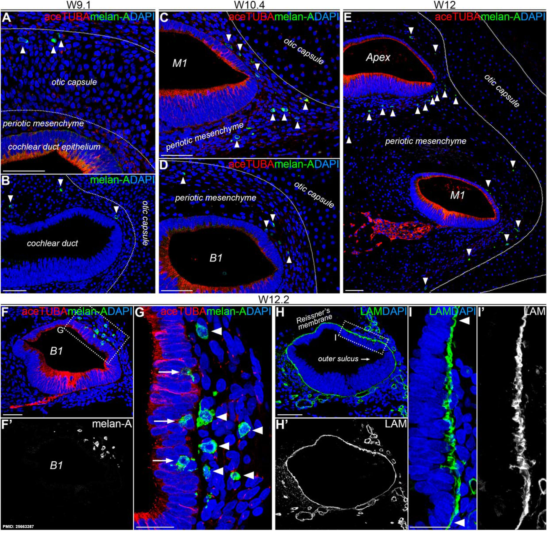 | 1,854 × 1,806 (754 KB) | Z8600021 (talk | contribs) | ==Migration of melanocytes into the stria vascularis of the human fetal cochlea== (A-B) A cochlea at W9.1 immunostained for acetylated tubulin (aceTUBA; red), melan-A (green) and DAPI (blue) (A) or melan-A (green) and DAPI (blue) (B). Arrowheads point... |
You cannot overwrite this file.
File usage
The following 7 pages use this file: