File:Human carnegie stage 3 label.jpg
From Embryology
No higher resolution available.
Human_carnegie_stage_3_label.jpg (500 × 377 pixels, file size: 26 KB, MIME type: image/jpeg)
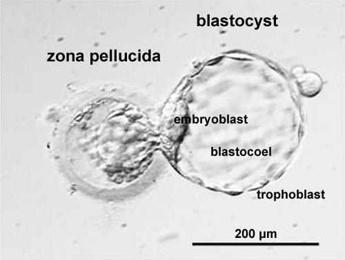
Human Embryo Carnegie stage 3
Human Blastocyst "hatching" from zona pellucida, in early Embryonic Development designated as Carnegie stage 3.
The zona pellucida is shown to the left of the image and the blastocyst is to the right of image.
Note:
- the small opening in the zona pellucida through which the blastocyst is hatching
- the flattened trophoblast cells forming the outer cell layer of the blastocyst
- the inner cell mass shown in the centre of the image and on the left-hand wall of the blastocyst
- the blastocoel forming a large fluid-filled space within the blastocyst
- Links: Carnegie stage 3 | Blastocyst Development | Week 1
Original image name: Figure 1 part b, Development of a blastomere-biopsied embryo into a hatching blastocyst. http://www.nature.com/nature/journal/v444/n7118/fig_tab/nature05142_F1.html (image extracted from full figure and adjusted in size and contrast)
Reference
<pubmed>16929302</pubmed>| Nature
File history
Click on a date/time to view the file as it appeared at that time.
| Date/Time | Thumbnail | Dimensions | User | Comment | |
|---|---|---|---|---|---|
| current | 13:08, 9 May 2011 | | 500 × 377 (26 KB) | S8600021 (talk | contribs) | ==Human Embryo Carnegie stage 3== Human Blastocyst "hatching" from zona pellucida, in early Embryonic Development designated as Carnegie stage 3. The '''zona pellucida''' is shown to the left of the image and the '''blastocyst''' is to the right |
You cannot overwrite this file.
File usage
The following 5 pages use this file: