File:Human- fetal week 10 sagittal plane D.jpg
Human-_fetal_week_10_sagittal_plane_D.jpg (500 × 573 pixels, file size: 105 KB, MIME type: image/jpeg)
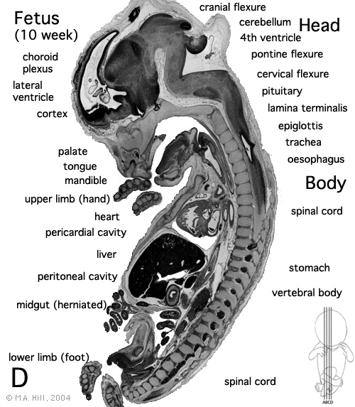
Human Fetus
female, 10 week, 40 mm CRL, early fetal, sagittal section, pelvic region
This stage of development is after the embryonic period (up to week 8) but still only 2 weeks into early fetal development.
Section D is the most midline of all sections. Planes A, B, C and D move towards the midline.
Original file name: H10wkUrogenAL.jpg http://embryology.med.unsw.edu.au/wwwhuman/Hum10wk/HumA.htm
Related Images
most lateral | lateral | medial | midline
Image Source: UNSW Embryology, no reproduction without permission.
File history
Click on a date/time to view the file as it appeared at that time.
| Date/Time | Thumbnail | Dimensions | User | Comment | |
|---|---|---|---|---|---|
| current | 14:21, 27 April 2010 | | 500 × 573 (105 KB) | S8600021 (talk | contribs) |
You cannot overwrite this file.
File usage
The following 15 pages use this file:
- 2010 BGD Practical 12 - Embryo to Fetus
- 2010 Lab 5
- 2011 Lab 12 - Embryo to Fetus
- 2011 Lab 5 - Fetal
- 2014 Group Project 3
- ANAT2341 Lab 11 - Embryo to Fetus
- ANAT2341 Lab 12 - Embryo to Fetus
- ANAT2341 Lab 5 - Fetal
- BGDA Practical 12 - Embryo to Fetus
- BGDB Gastrointestinal - Fetal
- BGD Lecture - Gastrointestinal System Development
- Fetal Development - 10 Weeks
- Lecture - Gastrointestinal Development
- Talk:2014 Group Project 3
- Talk:Foundations Practical - Week 9 to 36