File:Human- fetal week 10 sagittal plane D.jpg: Difference between revisions
From Embryology
No edit summary |
No edit summary |
||
| Line 12: | Line 12: | ||
==Related Images== | ==Related Images== | ||
[[:File:Human-_fetal_week_10_sagittal_plane_A.jpg|most lateral]] | [[:File:Human-_fetal_week_10_sagittal_plane_B.jpg|lateral]] | [[:File:Human-_fetal_week_10_sagittal_plane_C.jpg|medial]] | [[:File:Human-_fetal_week_10_sagittal_plane_D.jpg|midline]] | Human Fetus - [[:File:Human-_fetal_week_10_sagittal_plane_A.jpg|most lateral]] | [[:File:Human-_fetal_week_10_sagittal_plane_B.jpg|lateral]] | [[:File:Human-_fetal_week_10_sagittal_plane_C.jpg|medial]] | [[:File:Human-_fetal_week_10_sagittal_plane_D.jpg|midline]] | ||
Revision as of 15:26, 27 April 2010
Human Fetus
female, 10 week, 40 mm CRL, early fetal, sagittal section, pelvic region
This stage of development is after the embryonic period (up to week 8) but still only 2 weeks into early fetal development.
Section D is the most midline of all sections. Planes A, B, C and D move towards the midline.
Original file name: H10wkUrogenAL.jpg http://embryology.med.unsw.edu.au/wwwhuman/Hum10wk/HumA.htm
Related Images
Human Fetus - most lateral | lateral | medial | midline
Image Source: UNSW Embryology, no reproduction without permission.
File history
Click on a date/time to view the file as it appeared at that time.
| Date/Time | Thumbnail | Dimensions | User | Comment | |
|---|---|---|---|---|---|
| current | 14:21, 27 April 2010 | 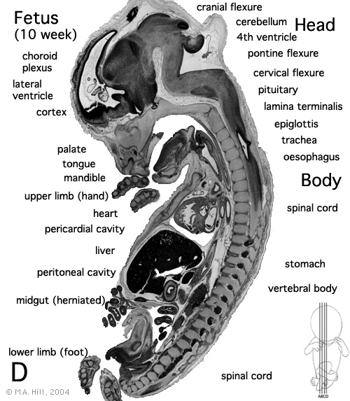 | 500 × 573 (105 KB) | S8600021 (talk | contribs) |
You cannot overwrite this file.
File usage
The following 15 pages use this file:
- 2010 BGD Practical 12 - Embryo to Fetus
- 2010 Lab 5
- 2011 Lab 12 - Embryo to Fetus
- 2011 Lab 5 - Fetal
- 2014 Group Project 3
- ANAT2341 Lab 11 - Embryo to Fetus
- ANAT2341 Lab 12 - Embryo to Fetus
- ANAT2341 Lab 5 - Fetal
- BGDA Practical 12 - Embryo to Fetus
- BGDB Gastrointestinal - Fetal
- BGD Lecture - Gastrointestinal System Development
- Fetal Development - 10 Weeks
- Lecture - Gastrointestinal Development
- Talk:2014 Group Project 3
- Talk:Foundations Practical - Week 9 to 36