File:Hughes1950 fig02.jpg

Original file (1,280 × 852 pixels, file size: 120 KB, MIME type: image/jpeg)
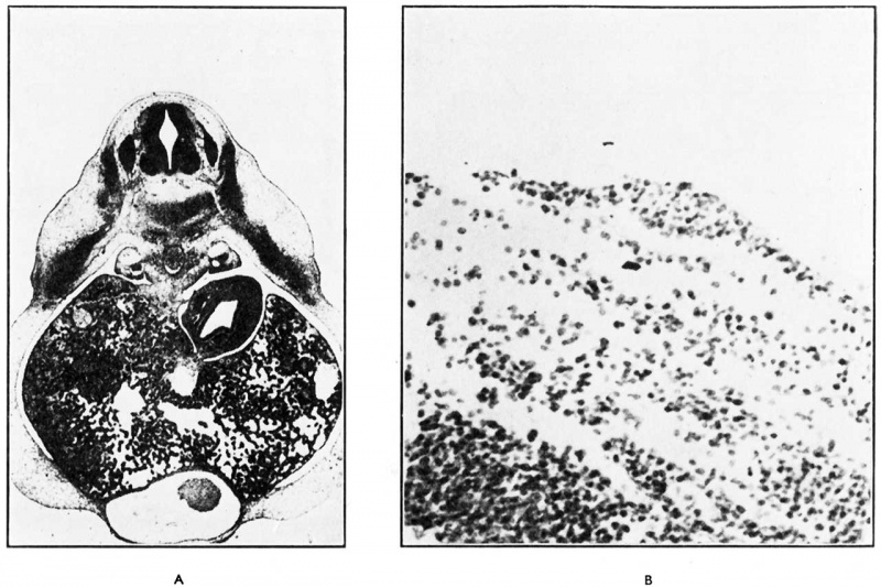
Fig. 2. Human Embryo 7 to 8 mm CRL
(A) The crest lies at the ventral end of the advancing dermatome in a dorso-lateral position ; it is surrounded by the mildly differentiated ectoderm of the “ mammary band.” There is a slight mesodermal condensation deep to the crest. (B) The cells forming the mammary crest possess a basal columnar layer with apical nuclei and clear basal cytoplasm. The remaining cells are polyhedral.
Reference
Hughes ES. Development of the mammary gland. (1950) Ann R Coll Surg Engl. 6(2):99-119. PMID 19309885
Cite this page: Hill, M.A. (2024, April 25) Embryology Hughes1950 fig02.jpg. Retrieved from https://embryology.med.unsw.edu.au/embryology/index.php/File:Hughes1950_fig02.jpg
- © Dr Mark Hill 2024, UNSW Embryology ISBN: 978 0 7334 2609 4 - UNSW CRICOS Provider Code No. 00098G
File history
Click on a date/time to view the file as it appeared at that time.
| Date/Time | Thumbnail | Dimensions | User | Comment | |
|---|---|---|---|---|---|
| current | 10:12, 15 August 2018 | | 1,280 × 852 (120 KB) | Z8600021 (talk | contribs) | |
| 10:12, 15 August 2018 | 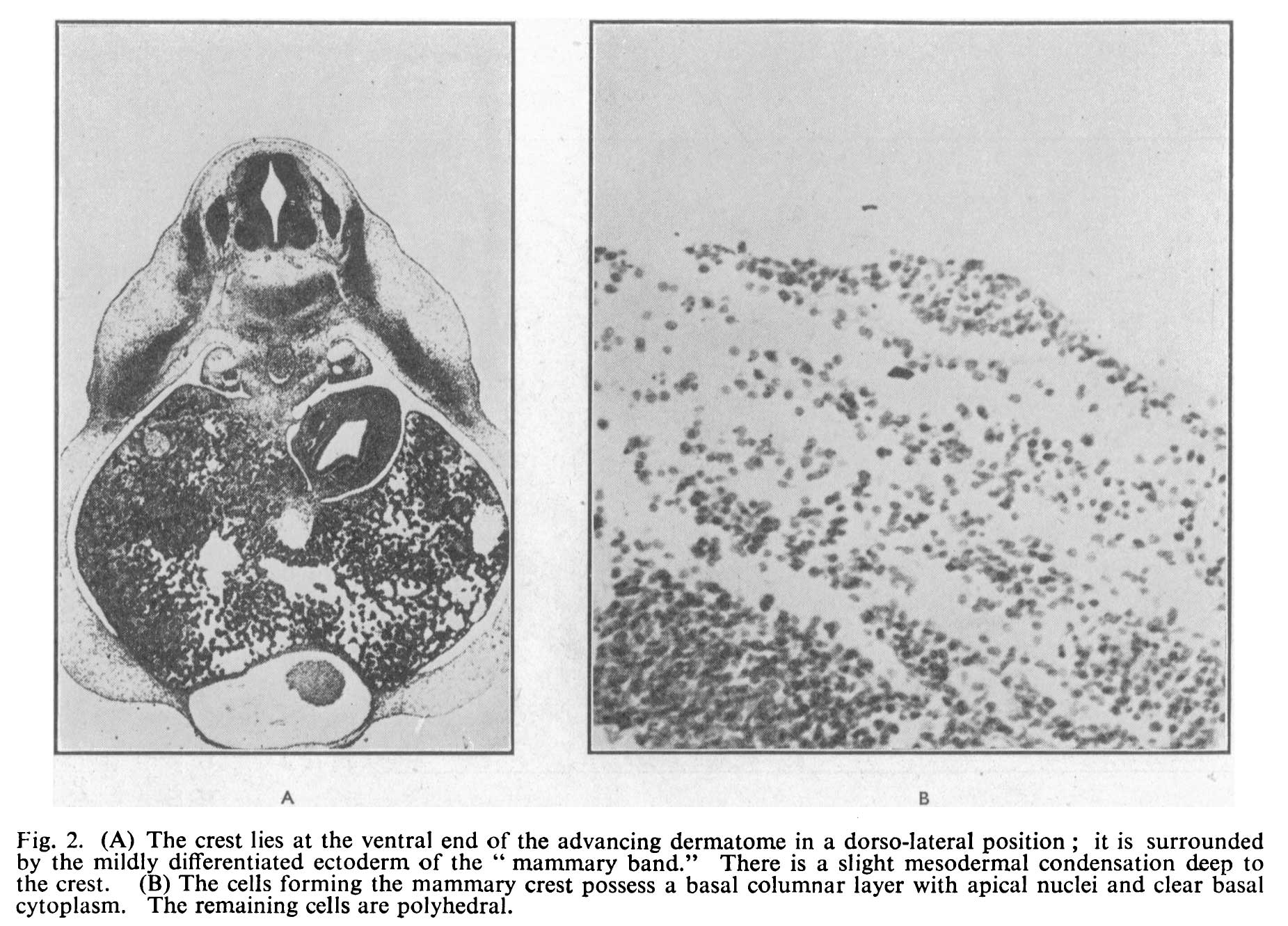 | 1,851 × 1,331 (279 KB) | Z8600021 (talk | contribs) | Fig. 2. (A) The crest lies at the ventral end of the advancing dermatome in a dorso-lateral position ; it is surrounded by the mildly differentiated ectoderm of the “ mammary band.” There is a slight mesodermal condensation deep to the crest. (B) T... |
You cannot overwrite this file.
File usage
The following page uses this file: