File:Huber1915 1fig18.jpg

Original file (622 × 800 pixels, file size: 40 KB, MIME type: image/jpeg)
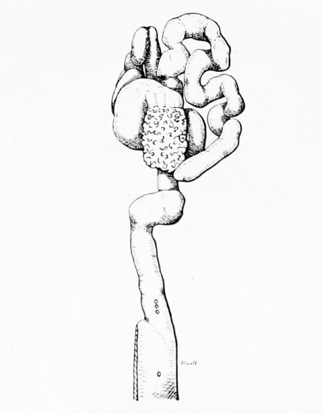
Fig. 18 Model of the left oviduct of rat
Rat No. 51, 4 days. X 10. A short segment of the upper end of the uterine horn was included in the reconstruction, lower end of the figure. The position of three of the morula masses, 12-cell to 16-cell stages, in the terminal part of the oviduct is to be noted, a further one is located in the upper part of the uterine horn. These are shown as if seen through a transparent wall. A fifth morula, situated in the uterine horn about 1.5 cm. from the entrance of the oviduct, is not included in the figure.
| Historic Disclaimer - information about historic embryology pages |
|---|
 |
- Albino Rat Links: Fig 14. Right Oviduct | Fig 15. 8 and 11-cell stages | The Development of the Albino Rat 1915
Cite this page: Hill, M.A. (2024, April 25) Embryology Huber1915 1fig18.jpg. Retrieved from https://embryology.med.unsw.edu.au/embryology/index.php/File:Huber1915_1fig18.jpg
- © Dr Mark Hill 2024, UNSW Embryology ISBN: 978 0 7334 2609 4 - UNSW CRICOS Provider Code No. 00098G
| Historic Disclaimer - information about historic embryology pages |
|---|
|
Cite this page: Hill, M.A. (2024, April 25) Embryology Huber1915 1fig18.jpg. Retrieved from https://embryology.med.unsw.edu.au/embryology/index.php/File:Huber1915_1fig18.jpg
- © Dr Mark Hill 2024, UNSW Embryology ISBN: 978 0 7334 2609 4 - UNSW CRICOS Provider Code No. 00098G
File history
Click on a date/time to view the file as it appeared at that time.
| Date/Time | Thumbnail | Dimensions | User | Comment | |
|---|---|---|---|---|---|
| current | 11:38, 7 April 2013 | | 622 × 800 (40 KB) | Z8600021 (talk | contribs) | ==Fig. 18 Model of the left oviduct of rat== Rat No. 51, 4 days. X 10. A short segment of the upper end of the uterine horn was included in the reconstruction, lower end of the figure. The position of three of the morula masses, 12-cell to 16-cell sta... |
You cannot overwrite this file.
File usage
The following page uses this file: