File:Huber1915 1fig03.jpg

From Embryology
Revision as of 10:36, 7 April 2013 by Z8600021 (talk | contribs)
(diff) ← Older revision | Latest revision (diff) | Newer revision → (diff)

Huber1915_1fig03.jpg(600 × 522 pixels, file size: 38 KB, MIME type: image/jpeg)

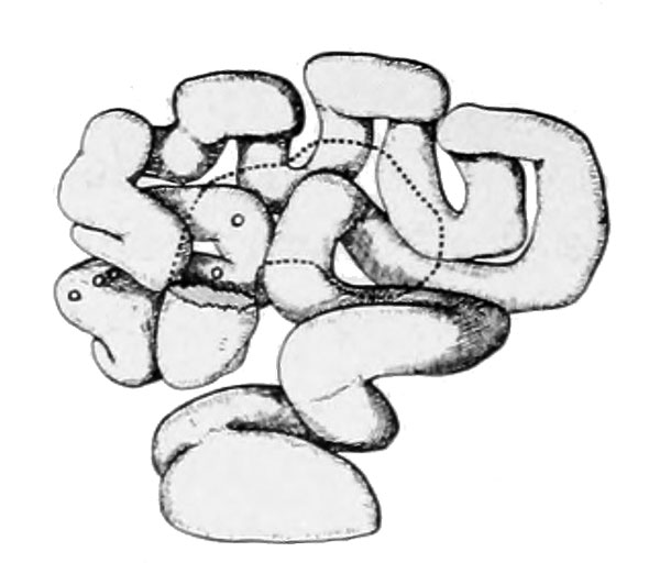
Fig. 3 Model of right oviduct of rat

Model of right oviduct of ratNo. 106, 24 hours. X 10. Fimbriated end and infundibulum removed in the drawing so as to expose underlying loops; their relative position given in dotted outline.

The position of the ova, which are outlined in circles, is shown as if seen through a transparent wall. The relative position of three of the eight ova found within this tube cannot be revealed in this view of the model.


Historic Disclaimer - information about historic embryology pages 
Mark Hill.jpg
Pages where the terms "Historic" (textbooks, papers, people, recommendations) appear on this site, and sections within pages where this disclaimer appears, indicate that the content and scientific understanding are specific to the time of publication. This means that while some scientific descriptions are still accurate, the terminology and interpretation of the developmental mechanisms reflect the understanding at the time of original publication and those of the preceding periods, these terms, interpretations and recommendations may not reflect our current scientific understanding.     (More? Embryology History | Historic Embryology Papers)
Albino Rat Links: Fig 14. Right Oviduct | Fig 15. 8 and 11-cell stages | The Development of the Albino Rat 1915


Normal: Introduction | Materials and Methods | Ovulation, Maturation and Fertilization | Pronuclear Stage | Segmentation Stages | 2-ceIl stage | 4-ceIl stage | 12 to 16-ceIl stages | Summary of segmentation stages | Completion of segmentation and blastodermic vesicle formation | Blastodermic vesicle | Late stages blastodermic vesicle | Egg-cylinder formation | Late stages in egg-cylinder | Conclusions | Literature cited | Figures
Abnormal: Introduction | Half Embryos in Mammalia | Degeneration of ova at the end of segmentation | Incomplete or retarded segmentation | Abnormal segmentation cavity formation | Degeneration of ova as a result of pathologic mucosa | Imperfect development of ectodermal vesicle | Two egg-cylinders in one decidual crypt | Conclusions | Literature cited


Cite this page: Hill, M.A. (2024, April 25) Embryology Huber1915 1fig03.jpg. Retrieved from https://embryology.med.unsw.edu.au/embryology/index.php/File:Huber1915_1fig03.jpg

What Links Here?
© Dr Mark Hill 2024, UNSW Embryology ISBN: 978 0 7334 2609 4 - UNSW CRICOS Provider Code No. 00098G


Normal: Introduction | Materials and Methods | Ovulation, Maturation and Fertilization | Pronuclear Stage | Segmentation Stages | 2-ceIl stage | 4-ceIl stage | 12 to 16-ceIl stages | Summary of segmentation stages | Completion of segmentation and blastodermic vesicle formation | Blastodermic vesicle | Late stages blastodermic vesicle | Egg-cylinder formation | Late stages in egg-cylinder | Conclusions | Literature cited | Figures
Abnormal: Introduction | Half Embryos in Mammalia | Degeneration of ova at the end of segmentation | Incomplete or retarded segmentation | Abnormal segmentation cavity formation | Degeneration of ova as a result of pathologic mucosa | Imperfect development of ectodermal vesicle | Two egg-cylinders in one decidual crypt | Conclusions | Literature cited
Historic Disclaimer - information about historic embryology pages 
Mark Hill.jpg
Pages where the terms "Historic" (textbooks, papers, people, recommendations) appear on this site, and sections within pages where this disclaimer appears, indicate that the content and scientific understanding are specific to the time of publication. This means that while some scientific descriptions are still accurate, the terminology and interpretation of the developmental mechanisms reflect the understanding at the time of original publication and those of the preceding periods, these terms, interpretations and recommendations may not reflect our current scientific understanding.     (More? Embryology History | Historic Embryology Papers)

Cite this page: Hill, M.A. (2024, April 25) Embryology Huber1915 1fig03.jpg. Retrieved from https://embryology.med.unsw.edu.au/embryology/index.php/File:Huber1915_1fig03.jpg

What Links Here?
© Dr Mark Hill 2024, UNSW Embryology ISBN: 978 0 7334 2609 4 - UNSW CRICOS Provider Code No. 00098G

File history

Click on a date/time to view the file as it appeared at that time.

Date/TimeThumbnailDimensionsUserComment
current10:35, 7 April 2013Thumbnail for version as of 10:35, 7 April 2013600 × 522 (38 KB)Z8600021 (talk | contribs)
00:22, 5 April 2013Thumbnail for version as of 00:22, 5 April 20131,000 × 507 (72 KB)Z8600021 (talk | contribs){{Huber1915 figures}} {{Huber1915_footer}}