File:HillH52 chorionic villi 08.jpg

Original file (1,200 × 900 pixels, file size: 229 KB, MIME type: image/jpeg)
Human Chorionic Villi
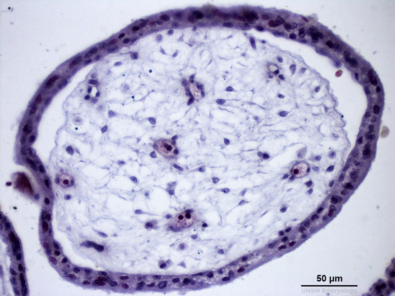
Human early placental villi development at 4-5 weeks. (Hill H52, x40) (Stain - Haematoxylin Eosin) scale bar - 50 μm)
Developing villi viewed in cross-section. Trophoblast shell enclosing mesenchyme (extra-embryonic mesoderm), containing embryonic blood vessels.
- Trophoblast layer consists of outer syncitiotrophoblast cells and inner cytotrophoblast cells.
- Gap between trophoblast and mesenchyme are shrinkage artefacts.
- H52 Links: Whole mount low power | Whole mount villi | Chorion and villi low power | Villi high power | Villi high power | Maternal decidua | Maternal decidua and Chorion | Villi cross-section
Image source: The images from the Hill Collection (part of the Embryological Collection) are reproduced with the permission of the Museum für Naturkunde, Leibniz Institute for Research on Evolution and Biodiversity. Images are for educational purposes only and must not be reproduced electronically or in writing without permission from the Museum für Naturkunde Berlin.
HillH52slide2x40_13.jpg 24092013 scaled to 1200px
File history
Click on a date/time to view the file as it appeared at that time.
| Date/Time | Thumbnail | Dimensions | User | Comment | |
|---|---|---|---|---|---|
| current | 15:39, 22 March 2014 |  | 1,200 × 900 (229 KB) | Z8600021 (talk | contribs) | ==Human Chorionic Villi== Human early placental villi development at 4-5 weeks. (Hill H52, x40) {{HE}} scale bar - 50 μm) {{HillH52}} HillH52slide2x40_13.jpg 24092013 scaled to 1200px |
You cannot overwrite this file.
File usage
The following page uses this file: