File:Hill1924 plate28-2.jpg

Original file (1,495 × 1,905 pixels, file size: 198 KB, MIME type: image/jpeg)
Plate 28
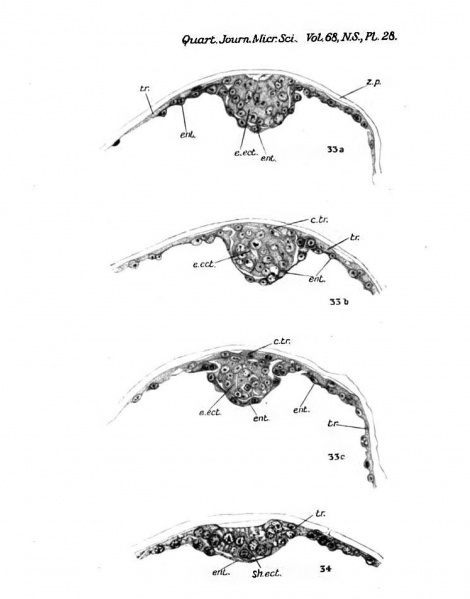
Figs. 33 a, 33 6, 33 c—Blastocyst 38, sections 1/2, 16/1, and 2/2 (x 400). In fig. 33 a, which passes through the central region of the embryonal ecto- derm (e.ect.), the covering trophoblast has disappeared and a slight depression is present on the surface of the ectodermal mass, the cells of which are now assuming a columnar arrangement. The covering trophobla3t (c.tr.) is still present over the more peripheral portion of the embryonal ectoderm (figs. 33 6 and 33 c).
Fig. 34.—Blastocyst 42. Section 7/2 ( x 420). sh.ect., shield-ectoderm.
File history
Click on a date/time to view the file as it appeared at that time.
| Date/Time | Thumbnail | Dimensions | User | Comment | |
|---|---|---|---|---|---|
| current | 16:29, 22 July 2015 |  | 1,495 × 1,905 (198 KB) | Z8600021 (talk | contribs) |
You cannot overwrite this file.
File usage
The following page uses this file: